 Movie
Movie Controller


+ Open data

- Basic information
| Entry | Database: PDB / ID: 6zn3 | |||||||||
|---|---|---|---|---|---|---|---|---|---|---|
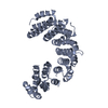
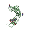
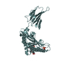
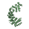
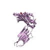
| Title | Plasmodium facliparum glideosome trimeric sub-complex | |||||||||
 Components Components |
| |||||||||
 Keywords Keywords | MOTOR PROTEIN / motility / glideosome / myosin / essential light chain | |||||||||
| Function / homology |  Function and homology information Function and homology informationpellicle / inner membrane pellicle complex / glideosome / myosin complex / myosin II complex / microfilament motor activity / cytoskeletal motor activity / actin filament organization / actin filament binding / actin cytoskeleton ...pellicle / inner membrane pellicle complex / glideosome / myosin complex / myosin II complex / microfilament motor activity / cytoskeletal motor activity / actin filament organization / actin filament binding / actin cytoskeleton / actin binding / calcium ion binding / ATP binding / membrane / plasma membrane / cytoplasm Similarity search - Function | |||||||||
| Biological species |  | |||||||||
| Method |  X-RAY DIFFRACTION / SYNCHROTRON / MOLECULAR REPLACEMENT / Resolution: 2.51 Å X-RAY DIFFRACTION / SYNCHROTRON / MOLECULAR REPLACEMENT / Resolution: 2.51 Å | |||||||||
 Authors Authors | Pazicky, S. / Loew, C. | |||||||||
| Funding support |  Sweden, Sweden,  Germany, 2items Germany, 2items
| |||||||||
 Citation Citation | Journal: Commun Biol / Year: 2020 Title: Structural role of essential light chains in the apicomplexan glideosome. Authors: Pazicky, S. / Dhamotharan, K. / Kaszuba, K. / Mertens, H.D.T. / Gilberger, T. / Svergun, D. / Kosinski, J. / Weininger, U. / Low, C. | |||||||||
| History |
|
- Structure visualization
| Structure viewer | Molecule: MolmilJmol/JSmol |
|---|
- Downloads & links
-Download
| PDBx/mmCIF format | 6zn3.cif.gz | 322.8 KB | Display | PDBx/mmCIF format |
|---|---|---|---|---|
| PDB format | pdb6zn3.ent.gz | 265.9 KB | Display | PDB format |
| PDBx/mmJSON format | 6zn3.json.gz | Tree view | PDBx/mmJSON format | |
| Others |  Other downloads Other downloads |
-Validation report
| Arichive directory | https://data.pdbj.org/pub/pdb/validation_reports/zn/6zn3ftp://data.pdbj.org/pub/pdb/validation_reports/zn/6zn3 | HTTPS FTP |
|---|
-Related structure data
| Related structure data | 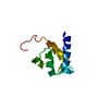 6tj3C 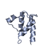 6tj4C  6tj5C  6tj6C  6tj7C 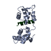 4aomS 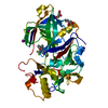 6jt4S S: Starting model for refinement C: citing same article ( |
|---|---|
| Similar structure data | |
| Other databases |
|


-Links
 PDBj
PDBj


- Assembly
| Deposited unit | 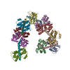
| ||||||||
|---|---|---|---|---|---|---|---|---|---|
| 1 | 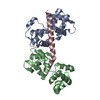
| ||||||||
| 2 | 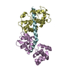
| ||||||||
| 3 | 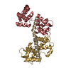
| ||||||||
| 4 | 
| ||||||||
| 5 | 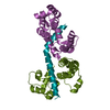
| ||||||||
| Unit cell |
|
-Components
| #1: Protein | Mass: 15779.875 Da / Num. of mol.: 5 Source method: isolated from a genetically manipulated source Source: (gene. exp.)  #2: Protein | Mass: 16788.572 Da / Num. of mol.: 5 Source method: isolated from a genetically manipulated source Source: (gene. exp.) #3: Protein/peptide | Mass: 5041.055 Da / Num. of mol.: 5 Source method: isolated from a genetically manipulated source Source: (gene. exp.) #4: Water | ChemComp-HOH / |  Mass: 18.015 Da / Num. of mol.: 3 / Source method: isolated from a natural source / Formula: H2O Mass: 18.015 Da / Num. of mol.: 3 / Source method: isolated from a natural source / Formula: H2O |
|---|
-Experimental details
-Experiment
| Experiment | Method: X-RAY DIFFRACTION / Number of used crystals: 1 |
|---|
- Sample preparation
| Crystal | Density Matthews: 4.58 Å3/Da / Density % sol: 73.13 % |
|---|---|
| Crystal grow | Temperature: 277.15 K / Method: vapor diffusion, sitting drop / pH: 6.5 Details: PEG 8000, ethylene glycol, di-ethyleneglycol, tri-ethyleneglycol, tetra-ethyleneglycol, penta-ethyleneglycol, imidazole, MES |
-Data collection
| Diffraction | Mean temperature: 80 K / Serial crystal experiment: N |
|---|---|
| Diffraction source | Source: SYNCHROTRON / Site: PETRA III, EMBL c/o DESY / Beamline: P13 (MX1) / Wavelength: 0.9793 Å |
| Detector | Type: DECTRIS PILATUS 6M / Detector: PIXEL / Date: May 19, 2020 |
| Radiation | Protocol: SINGLE WAVELENGTH / Monochromatic (M) / Laue (L): M / Scattering type: x-ray |
| Radiation wavelength | Wavelength: 0.9793 Å / Relative weight: 1 |
| Reflection | Resolution: 2.51→47.42 Å / Num. obs: 114354 / % possible obs: 83.53 % / Redundancy: 6.7 % / Biso Wilson estimate: 81.46 Å2 / CC1/2: 0.998 / CC star: 1 / Rmerge(I) obs: 0.0874 / Rpim(I) all: 0.03701 / Rrim(I) all: 0.09505 / Net I/σ(I): 12.69 |
| Reflection shell | Resolution: 2.51→2.604 Å / Redundancy: 6.6 % / Rmerge(I) obs: 3.79 / Mean I/σ(I) obs: 0.55 / Num. unique obs: 911 / CC1/2: 0.111 / CC star: 0.448 / Rpim(I) all: 1.602 / Rrim(I) all: 4.118 / % possible all: 7.97 |
- Processing
| Software |
| ||||||||||||||||||||
|---|---|---|---|---|---|---|---|---|---|---|---|---|---|---|---|---|---|---|---|---|---|
| Refinement | Method to determine structure: MOLECULAR REPLACEMENT Starting model: 6jt4, 4aom Resolution: 2.51→47.42 Å / Cor.coef. Fo:Fc: 0.958 / Cor.coef. Fo:Fc free: 0.941 / SU B: 9.812 / SU ML: 0.201 / Cross valid method: THROUGHOUT / σ(F): 2.51 / ESU R: 0.314 / ESU R Free: 0.244 / Stereochemistry target values: MAXIMUM LIKELIHOOD / Details: HYDROGENS HAVE BEEN ADDED IN THE RIDING POSITIONS
| ||||||||||||||||||||
| Solvent computation | Ion probe radii: 0.8 Å / Shrinkage radii: 0.8 Å / VDW probe radii: 1.2 Å / Solvent model: MASK | ||||||||||||||||||||
| Displacement parameters | Biso mean: 91.468 Å2
| ||||||||||||||||||||
| Refinement step | Cycle: 1 / Resolution: 2.51→47.42 Å
| ||||||||||||||||||||
| Refine LS restraints | Type: r_bond_refined_d / Dev ideal: 0.009 / Dev ideal target: 0.013 / Number: 13175 | ||||||||||||||||||||
| LS refinement shell | Resolution: 2.514→2.579 Å / Total num. of bins used: 20
|
