 Movie
Movie Controller


+ Open data

- Basic information
| Entry | Database: PDB / ID: 6tj6 | |||||||||
|---|---|---|---|---|---|---|---|---|---|---|
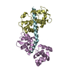
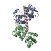
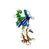
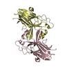
| Title | T. gondii myosin A trimeric complex with ELC1, calcium-free | |||||||||
 Components Components |
| |||||||||
 Keywords Keywords | MOTOR PROTEIN / motility / glideosome / light chain / myosin | |||||||||
| Function / homology |  Function and homology information Function and homology informationmyosin complex / myosin II complex / microfilament motor activity / cytoskeletal motor activity / actin filament organization / actin filament binding / actin binding / calcium ion binding / ATP binding / plasma membrane / cytoplasm Similarity search - Function | |||||||||
| Biological species |  | |||||||||
| Method |  X-RAY DIFFRACTION / SYNCHROTRON / MOLECULAR REPLACEMENT / Resolution: 2 Å X-RAY DIFFRACTION / SYNCHROTRON / MOLECULAR REPLACEMENT / Resolution: 2 Å | |||||||||
 Authors Authors | Pazicky, S. / Loew, C. | |||||||||
| Funding support |  Germany, Germany,  Sweden, 2items Sweden, 2items
| |||||||||
 Citation Citation | Journal: Commun Biol / Year: 2020 Title: Structural role of essential light chains in the apicomplexan glideosome. Authors: Pazicky, S. / Dhamotharan, K. / Kaszuba, K. / Mertens, H.D.T. / Gilberger, T. / Svergun, D. / Kosinski, J. / Weininger, U. / Low, C. | |||||||||
| History |
|
- Structure visualization
| Structure viewer | Molecule: MolmilJmol/JSmol |
|---|
- Downloads & links
-Download
| PDBx/mmCIF format | 6tj6.cif.gz | 172.3 KB | Display | PDBx/mmCIF format |
|---|---|---|---|---|
| PDB format | pdb6tj6.ent.gz | 113.9 KB | Display | PDB format |
| PDBx/mmJSON format | 6tj6.json.gz | Tree view | PDBx/mmJSON format | |
| Others |  Other downloads Other downloads |
-Validation report
| Arichive directory | https://data.pdbj.org/pub/pdb/validation_reports/tj/6tj6ftp://data.pdbj.org/pub/pdb/validation_reports/tj/6tj6 | HTTPS FTP |
|---|
-Related structure data
| Related structure data | 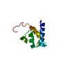 6tj3C 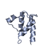 6tj4C  6tj5C  6tj7C  6zn3C  5vt9S S: Starting model for refinement C: citing same article ( |
|---|---|
| Similar structure data |


-Links
 PDBj
PDBj


- Assembly
| Deposited unit | 
| ||||||||||||
|---|---|---|---|---|---|---|---|---|---|---|---|---|---|
| 1 |
| ||||||||||||
| Unit cell |
|
-Components
| #1: Protein | Mass: 14881.957 Da / Num. of mol.: 1 Source method: isolated from a genetically manipulated source Source: (gene. exp.)  |
|---|---|
| #2: Protein | Mass: 17198.916 Da / Num. of mol.: 1 Source method: isolated from a genetically manipulated source Source: (gene. exp.) |
| #3: Protein/peptide | Mass: 5340.347 Da / Num. of mol.: 1 Source method: isolated from a genetically manipulated source Source: (gene. exp.) |
| #4: Chemical | ChemComp-IMD /   Mass: 69.085 Da / Num. of mol.: 1 / Source method: obtained synthetically / Formula: C3H5N2 Mass: 69.085 Da / Num. of mol.: 1 / Source method: obtained synthetically / Formula: C3H5N2 |
| #5: Water | ChemComp-HOH /  Mass: 18.015 Da / Num. of mol.: 147 / Source method: isolated from a natural source / Formula: H2O Mass: 18.015 Da / Num. of mol.: 147 / Source method: isolated from a natural source / Formula: H2O |
| Has ligand of interest | N |
-Experimental details
-Experiment
| Experiment | Method: X-RAY DIFFRACTION / Number of used crystals: 1 |
|---|
- Sample preparation
| Crystal | Density Matthews: 2.67 Å3/Da / Density % sol: 54 % |
|---|---|
| Crystal grow | Temperature: 292.15 K / Method: vapor diffusion, sitting drop / pH: 7 / Details: PEG 8000, Tris, Lithium Chloride |
-Data collection
| Diffraction | Mean temperature: 100 K / Serial crystal experiment: N |
|---|---|
| Diffraction source | Source: SYNCHROTRON / Site: PETRA III, EMBL c/o DESY / Beamline: P13 (MX1) / Wavelength: 0.9762 Å |
| Detector | Type: DECTRIS PILATUS 6M / Detector: PIXEL / Date: Nov 15, 2019 |
| Radiation | Protocol: SINGLE WAVELENGTH / Monochromatic (M) / Laue (L): M / Scattering type: x-ray |
| Radiation wavelength | Wavelength: 0.9762 Å / Relative weight: 1 |
| Reflection | Resolution: 2→40.9 Å / Num. obs: 27763 / % possible obs: 99.91 % / Redundancy: 13.5 % / Biso Wilson estimate: 48.32 Å2 / CC1/2: 0.99 / Rmerge(I) obs: 0.04 / Rpim(I) all: 0.01 / Rrim(I) all: 0.15 / Net I/σ(I): 31 |
| Reflection shell | Resolution: 2→2.07 Å / Redundancy: 12.9 % / Rmerge(I) obs: 1.35 / Mean I/σ(I) obs: 1.74 / Num. unique obs: 2711 / CC1/2: 0.72 / Rpim(I) all: 0.39 / Rrim(I) all: 1.41 / % possible all: 99.38 |
- Processing
| Software |
| |||||||||||||||||||||||||||||||||||||||||||||||||||||||||||||||||||||||||||||
|---|---|---|---|---|---|---|---|---|---|---|---|---|---|---|---|---|---|---|---|---|---|---|---|---|---|---|---|---|---|---|---|---|---|---|---|---|---|---|---|---|---|---|---|---|---|---|---|---|---|---|---|---|---|---|---|---|---|---|---|---|---|---|---|---|---|---|---|---|---|---|---|---|---|---|---|---|---|---|
| Refinement | Method to determine structure: MOLECULAR REPLACEMENT Starting model: 5vt9 Resolution: 2→40.9 Å / SU ML: 0.2589 / Cross valid method: FREE R-VALUE / σ(F): 1.35 / Phase error: 25.3194
| |||||||||||||||||||||||||||||||||||||||||||||||||||||||||||||||||||||||||||||
| Solvent computation | Shrinkage radii: 0.9 Å / VDW probe radii: 1.11 Å | |||||||||||||||||||||||||||||||||||||||||||||||||||||||||||||||||||||||||||||
| Displacement parameters | Biso mean: 65.41 Å2 | |||||||||||||||||||||||||||||||||||||||||||||||||||||||||||||||||||||||||||||
| Refinement step | Cycle: LAST / Resolution: 2→40.9 Å
| |||||||||||||||||||||||||||||||||||||||||||||||||||||||||||||||||||||||||||||
| Refine LS restraints |
| |||||||||||||||||||||||||||||||||||||||||||||||||||||||||||||||||||||||||||||
| LS refinement shell |
| |||||||||||||||||||||||||||||||||||||||||||||||||||||||||||||||||||||||||||||
| Refinement TLS params. | Method: refined / Origin x: -14.3905764905 Å / Origin y: -43.6774644172 Å / Origin z: -11.0030210361 Å
| |||||||||||||||||||||||||||||||||||||||||||||||||||||||||||||||||||||||||||||
| Refinement TLS group | Selection details: all |
