 Movie
Movie Controller

[English] 日本語
 Yorodumi
- PDB-3qbz: Crystal structure of the Rad53-recognition domain of Saccharomyce... -
+ Open data

- Basic information
| Entry | Database: PDB / ID: 3qbz | ||||||
|---|---|---|---|---|---|---|---|
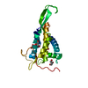
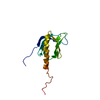
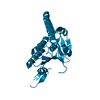
| Title | Crystal structure of the Rad53-recognition domain of Saccharomyces cerevisiae Dbf4 | ||||||
 Components Components | DDK kinase regulatory subunit DBF4 | ||||||
 Keywords Keywords | CELL CYCLE / FHA domain / Rad53 / replication checkpoint | ||||||
| Function / homology |  Function and homology information Function and homology informationpositive regulation of DNA replication initiation / negative regulation of exit from mitosis / Dbf4-dependent protein kinase complex / positive regulation of meiosis I / positive regulation of nuclear cell cycle DNA replication / regulation of cell cycle phase transition / premeiotic DNA replication / Activation of the pre-replicative complex / Activation of ATR in response to replication stress / mitotic DNA replication checkpoint signaling ...positive regulation of DNA replication initiation / negative regulation of exit from mitosis / Dbf4-dependent protein kinase complex / positive regulation of meiosis I / positive regulation of nuclear cell cycle DNA replication / regulation of cell cycle phase transition / premeiotic DNA replication / Activation of the pre-replicative complex / Activation of ATR in response to replication stress / mitotic DNA replication checkpoint signaling / mitotic intra-S DNA damage checkpoint signaling / DNA replication origin binding / chromosome, centromeric region / DNA replication initiation / protein serine/threonine kinase activator activity / chromosome segregation / cell division / centrosome / chromatin / zinc ion binding / nucleus / cytoplasm Similarity search - Function | ||||||
| Biological species |  | ||||||
| Method |  X-RAY DIFFRACTION / SYNCHROTRON / MOLECULAR REPLACEMENT / Resolution: 2.692 Å X-RAY DIFFRACTION / SYNCHROTRON / MOLECULAR REPLACEMENT / Resolution: 2.692 Å | ||||||
 Authors Authors | Matthews, L.A. / Jones, D.R. / Prasad, A.A. / Duncker, B.P. / Guarne, A. | ||||||
 Citation Citation | Journal: J.Biol.Chem. / Year: 2012 Title: Saccharomyces cerevisiae Dbf4 has unique fold necessary for interaction with Rad53 kinase. Authors: Matthews, L.A. / Jones, D.R. / Prasad, A.A. / Duncker, B.P. / Guarne, A. #1: Journal: Acta Crystallogr.,Sect.F / Year: 2009Title: Crystallization and preliminary X-ray diffraction analysis of motif N from Saccharomyces cerevisiae Dbf4 Authors: Matthews, L.A. / Duong, A. / Prasad, A.A. / Duncker, B.P. / Guarne, A. | ||||||
| History |
|
- Structure visualization
| Structure viewer | Molecule: MolmilJmol/JSmol |
|---|
- Downloads & links
-Download
| PDBx/mmCIF format | 3qbz.cif.gz | 68.4 KB | Display | PDBx/mmCIF format |
|---|---|---|---|---|
| PDB format | pdb3qbz.ent.gz | 50.8 KB | Display | PDB format |
| PDBx/mmJSON format | 3qbz.json.gz | Tree view | PDBx/mmJSON format | |
| Others |  Other downloads Other downloads |
-Validation report
| Arichive directory | https://data.pdbj.org/pub/pdb/validation_reports/qb/3qbzftp://data.pdbj.org/pub/pdb/validation_reports/qb/3qbz | HTTPS FTP |
|---|
-Related structure data
| Related structure data |  3oq4S S: Starting model for refinement |
|---|---|
| Similar structure data |


-Links
 PDBj
PDBj


- Assembly
| Deposited unit | 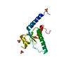
| ||||||||
|---|---|---|---|---|---|---|---|---|---|
| 1 |
| ||||||||
| 2 | 
| ||||||||
| Unit cell |
|
-Components
| #1: Protein | Mass: 18993.949 Da / Num. of mol.: 1 / Fragment: residues 66-221, HBRCT domain Source method: isolated from a genetically manipulated source Details: Dbf4 residues 66-221 subcloned in pET15b using NdeI-BamHI Source: (gene. exp.) Gene: D4205, DBF4, DNA52, YD9609.07C, YDR052C / Plasmid: pET15b / Production host:  | ||
|---|---|---|---|
| #2: Chemical |   Mass: 96.063 Da / Num. of mol.: 3 / Source method: obtained synthetically / Formula: SO4 Mass: 96.063 Da / Num. of mol.: 3 / Source method: obtained synthetically / Formula: SO4#3: Water | ChemComp-HOH / |  Mass: 18.015 Da / Num. of mol.: 10 / Source method: isolated from a natural source / Formula: H2O Mass: 18.015 Da / Num. of mol.: 10 / Source method: isolated from a natural source / Formula: H2O |
-Experimental details
-Experiment
| Experiment | Method: X-RAY DIFFRACTION / Number of used crystals: 1 |
|---|
- Sample preparation
| Crystal | Density Matthews: 2.76 Å3/Da / Density % sol: 55.47 % |
|---|---|
| Crystal grow | Temperature: 277 K / Method: vapor diffusion, hanging drop / pH: 6.5 Details: 1.9 M ammonium sulfate 50 mM sodium cacodylate 15 mM CYMAL-7, pH 6.5, VAPOR DIFFUSION, HANGING DROP, temperature 277K |
-Data collection
| Diffraction | Mean temperature: 100 K |
|---|---|
| Diffraction source | Source: SYNCHROTRON / Site: NSLS  / Beamline: X25 / Wavelength: 1.1 Å / Beamline: X25 / Wavelength: 1.1 Å |
| Detector | Type: ADSC QUANTUM 315 / Detector: CCD / Date: Nov 10, 2010 / Details: mirrors |
| Radiation | Monochromator: Si(111) / Protocol: SINGLE WAVELENGTH / Monochromatic (M) / Laue (L): M / Scattering type: x-ray |
| Radiation wavelength | Wavelength: 1.1 Å / Relative weight: 1 |
| Reflection | Resolution: 2.69→50 Å / Num. obs: 6371 / % possible obs: 99.7 % / Observed criterion σ(F): 0 / Observed criterion σ(I): 0 / Redundancy: 6.9 % / Biso Wilson estimate: 53.46 Å2 / Rmerge(I) obs: 0.057 / Rsym value: 0.042 / Net I/σ(I): 31 |
| Reflection shell | Resolution: 2.69→2.74 Å / Redundancy: 6.2 % / Rmerge(I) obs: 0.419 / Mean I/σ(I) obs: 3.5 / Num. unique all: 298 / Rsym value: 0.344 / % possible all: 99.7 |
- Processing
| Software |
| |||||||||||||||||||||||||||||||||||||||||||||||||||||||||||||||||||||||||||||||||||||||||||||||||||||||||||||||||||||||||||||||||||||||||||||||||||||||||||||||||||||||||||||||||||||||||||||||||||||||||||||||||||||||||||||||||||||||||||||||||||||||||||||||||||||||||||||||||||
|---|---|---|---|---|---|---|---|---|---|---|---|---|---|---|---|---|---|---|---|---|---|---|---|---|---|---|---|---|---|---|---|---|---|---|---|---|---|---|---|---|---|---|---|---|---|---|---|---|---|---|---|---|---|---|---|---|---|---|---|---|---|---|---|---|---|---|---|---|---|---|---|---|---|---|---|---|---|---|---|---|---|---|---|---|---|---|---|---|---|---|---|---|---|---|---|---|---|---|---|---|---|---|---|---|---|---|---|---|---|---|---|---|---|---|---|---|---|---|---|---|---|---|---|---|---|---|---|---|---|---|---|---|---|---|---|---|---|---|---|---|---|---|---|---|---|---|---|---|---|---|---|---|---|---|---|---|---|---|---|---|---|---|---|---|---|---|---|---|---|---|---|---|---|---|---|---|---|---|---|---|---|---|---|---|---|---|---|---|---|---|---|---|---|---|---|---|---|---|---|---|---|---|---|---|---|---|---|---|---|---|---|---|---|---|---|---|---|---|---|---|---|---|---|---|---|---|---|---|---|---|---|---|---|---|---|---|---|---|---|---|---|---|---|---|---|---|---|---|---|---|---|---|---|---|---|---|---|---|---|---|---|---|---|---|---|---|---|---|---|---|---|---|---|---|---|---|
| Refinement | Method to determine structure: MOLECULAR REPLACEMENT Starting model: PDB entry 3OQ4 Resolution: 2.692→38.823 Å / SU ML: 0.43 / Cross valid method: THROUGHOUT / σ(F): 0 / σ(I): 0 / Stereochemistry target values: MLHL
| |||||||||||||||||||||||||||||||||||||||||||||||||||||||||||||||||||||||||||||||||||||||||||||||||||||||||||||||||||||||||||||||||||||||||||||||||||||||||||||||||||||||||||||||||||||||||||||||||||||||||||||||||||||||||||||||||||||||||||||||||||||||||||||||||||||||||||||||||||
| Solvent computation | Shrinkage radii: 0.83 Å / VDW probe radii: 1.1 Å / Solvent model: FLAT BULK SOLVENT MODEL / Bsol: 42.732 Å2 / ksol: 0.315 e/Å3 | |||||||||||||||||||||||||||||||||||||||||||||||||||||||||||||||||||||||||||||||||||||||||||||||||||||||||||||||||||||||||||||||||||||||||||||||||||||||||||||||||||||||||||||||||||||||||||||||||||||||||||||||||||||||||||||||||||||||||||||||||||||||||||||||||||||||||||||||||||
| Displacement parameters |
| |||||||||||||||||||||||||||||||||||||||||||||||||||||||||||||||||||||||||||||||||||||||||||||||||||||||||||||||||||||||||||||||||||||||||||||||||||||||||||||||||||||||||||||||||||||||||||||||||||||||||||||||||||||||||||||||||||||||||||||||||||||||||||||||||||||||||||||||||||
| Refinement step | Cycle: LAST / Resolution: 2.692→38.823 Å
| |||||||||||||||||||||||||||||||||||||||||||||||||||||||||||||||||||||||||||||||||||||||||||||||||||||||||||||||||||||||||||||||||||||||||||||||||||||||||||||||||||||||||||||||||||||||||||||||||||||||||||||||||||||||||||||||||||||||||||||||||||||||||||||||||||||||||||||||||||
| Refine LS restraints |
| |||||||||||||||||||||||||||||||||||||||||||||||||||||||||||||||||||||||||||||||||||||||||||||||||||||||||||||||||||||||||||||||||||||||||||||||||||||||||||||||||||||||||||||||||||||||||||||||||||||||||||||||||||||||||||||||||||||||||||||||||||||||||||||||||||||||||||||||||||
| LS refinement shell | Refine-ID: X-RAY DIFFRACTION / % reflection obs: 100 %
| |||||||||||||||||||||||||||||||||||||||||||||||||||||||||||||||||||||||||||||||||||||||||||||||||||||||||||||||||||||||||||||||||||||||||||||||||||||||||||||||||||||||||||||||||||||||||||||||||||||||||||||||||||||||||||||||||||||||||||||||||||||||||||||||||||||||||||||||||||
| Refinement TLS params. | Method: refined / Refine-ID: X-RAY DIFFRACTION
| |||||||||||||||||||||||||||||||||||||||||||||||||||||||||||||||||||||||||||||||||||||||||||||||||||||||||||||||||||||||||||||||||||||||||||||||||||||||||||||||||||||||||||||||||||||||||||||||||||||||||||||||||||||||||||||||||||||||||||||||||||||||||||||||||||||||||||||||||||
| Refinement TLS group |
|
