 Movie
Movie Controller

[English] 日本語
 Yorodumi
- PDB-5t2s: Structure of the FHA1 domain of Rad53 bound simultaneously to the... -
+ Open data

- Basic information
| Entry | Database: PDB / ID: 5t2s | ||||||
|---|---|---|---|---|---|---|---|
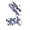
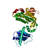
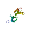
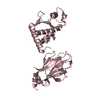
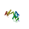
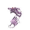
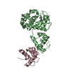
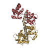
| Title | Structure of the FHA1 domain of Rad53 bound simultaneously to the BRCT domain of Dbf4 and a phosphopeptide. | ||||||
 Components Components |
| ||||||
 Keywords Keywords | CELL CYCLE / FHA / BRCT / phosphopeptide / protein chimera | ||||||
| Function / homology |  Function and homology information Function and homology informationpositive regulation of spindle attachment to meiosis I kinetochore / positive regulation of meiotic DNA double-strand break formation involved in reciprocal meiotic recombination / positive regulation of DNA replication initiation / negative regulation of exit from mitosis / G2/M DNA damage checkpoint / Dbf4-dependent protein kinase complex / Chk1/Chk2(Cds1) mediated inactivation of Cyclin B:Cdk1 complex / deoxyribonucleoside triphosphate biosynthetic process / positive regulation of protein localization to kinetochore / positive regulation of meiosis I ...positive regulation of spindle attachment to meiosis I kinetochore / positive regulation of meiotic DNA double-strand break formation involved in reciprocal meiotic recombination / positive regulation of DNA replication initiation / negative regulation of exit from mitosis / G2/M DNA damage checkpoint / Dbf4-dependent protein kinase complex / Chk1/Chk2(Cds1) mediated inactivation of Cyclin B:Cdk1 complex / deoxyribonucleoside triphosphate biosynthetic process / positive regulation of protein localization to kinetochore / positive regulation of meiosis I / positive regulation of nuclear cell cycle DNA replication / regulation of cell cycle phase transition / mitotic DNA damage checkpoint signaling / negative regulation of phosphorylation / mitotic DNA replication preinitiation complex assembly / premeiotic DNA replication / Activation of the pre-replicative complex / Activation of ATR in response to replication stress / dual-specificity kinase / Recruitment and ATM-mediated phosphorylation of repair and signaling proteins at DNA double strand breaks / mitotic DNA replication checkpoint signaling / double-strand break repair via break-induced replication / mitotic intra-S DNA damage checkpoint signaling / protein-containing complex localization / Ubiquitin-Mediated Degradation of Phosphorylated Cdc25A / telomere maintenance in response to DNA damage / negative regulation of DNA damage checkpoint / DNA replication origin binding / chromosome, centromeric region / DNA replication initiation / regulation of DNA repair / protein serine/threonine/tyrosine kinase activity / DNA damage checkpoint signaling / protein serine/threonine kinase activator activity / chromosome segregation / intracellular protein localization / protein tyrosine kinase activity / protein kinase activity / non-specific serine/threonine protein kinase / protein serine kinase activity / cell division / DNA repair / protein serine/threonine kinase activity / centrosome / chromatin / signal transduction / zinc ion binding / ATP binding / metal ion binding / identical protein binding / nucleus / cytosol / cytoplasm Similarity search - Function | ||||||
| Biological species |  | ||||||
| Method |  X-RAY DIFFRACTION / SYNCHROTRON / MOLECULAR REPLACEMENT / Resolution: 2.4 Å X-RAY DIFFRACTION / SYNCHROTRON / MOLECULAR REPLACEMENT / Resolution: 2.4 Å | ||||||
 Authors Authors | Guarne, A. / Almawi, A. / Matthews, L. | ||||||
| Funding support |  Canada, 1items Canada, 1items
| ||||||
 Citation Citation | Journal: Sci Rep / Year: 2016 Title: 'AND' logic gates at work: Crystal structure of Rad53 bound to Dbf4 and Cdc7. Authors: Almawi, A.W. / Matthews, L.A. / Myrox, P. / Boulton, S. / Lai, C. / Moraes, T. / Melacini, G. / Ghirlando, R. / Duncker, B.P. / Guarne, A. | ||||||
| History |
|
- Structure visualization
| Structure viewer | Molecule: MolmilJmol/JSmol |
|---|
- Downloads & links
-Download
| PDBx/mmCIF format | 5t2s.cif.gz | 120.7 KB | Display | PDBx/mmCIF format |
|---|---|---|---|---|
| PDB format | pdb5t2s.ent.gz | 91.9 KB | Display | PDB format |
| PDBx/mmJSON format | 5t2s.json.gz | Tree view | PDBx/mmJSON format | |
| Others |  Other downloads Other downloads |
-Validation report
| Arichive directory | https://data.pdbj.org/pub/pdb/validation_reports/t2/5t2sftp://data.pdbj.org/pub/pdb/validation_reports/t2/5t2s | HTTPS FTP |
|---|
-Related structure data
| Related structure data |  5t2fC  1g6gS  3qbzS C: citing same article ( S: Starting model for refinement |
|---|---|
| Similar structure data |
-Links
 PDBj
PDBj


- Assembly
| Deposited unit | 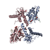
| ||||||||
|---|---|---|---|---|---|---|---|---|---|
| 1 | 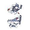
| ||||||||
| 2 | 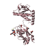
| ||||||||
| 3 |
| ||||||||
| Unit cell |
|
-Components
| #1: Protein | Mass: 30571.002 Da / Num. of mol.: 2 Fragment: UNP P32325 residues 105-220 linked to UNP P22216 residues 22-162 via LINKER residues VDGS Source method: isolated from a genetically manipulated source Source: (gene. exp.) Strain: ATCC 204508 / S288c Gene: DBF4, DNA52, YDR052C, D4205, YD9609.07C, RAD53, MEC2, SAD1, SPK1, YPL153C, P2588 Plasmid: modified pET15b Details (production host): Contains aTEV-removable His6-SUMO tag Production host:  References: UniProt: P32325, UniProt: P22216, dual-specificity kinase #2: Protein/peptide | Mass: 1347.147 Da / Num. of mol.: 2 / Source method: obtained synthetically / Details: short phosphorylated peptide derived from Cdc7 / Source: (synth.) #3: Chemical | ChemComp-GOL /   Mass: 92.094 Da / Num. of mol.: 11 / Source method: obtained synthetically / Formula: C3H8O3 Mass: 92.094 Da / Num. of mol.: 11 / Source method: obtained synthetically / Formula: C3H8O3#4: Water | ChemComp-HOH / |  Mass: 18.015 Da / Num. of mol.: 177 / Source method: isolated from a natural source / Formula: H2O Mass: 18.015 Da / Num. of mol.: 177 / Source method: isolated from a natural source / Formula: H2OHas protein modification | Y | |
|---|
-Experimental details
-Experiment
| Experiment | Method: X-RAY DIFFRACTION / Number of used crystals: 1 |
|---|
- Sample preparation
| Crystal | Density Matthews: 2.91 Å3/Da / Density % sol: 57.73 % |
|---|---|
| Crystal grow | Temperature: 277 K / Method: vapor diffusion, sitting drop / pH: 8.5 / Details: 100 mM TRIS pH 8.5 12.5 % PEG 3350 (v/v) / PH range: 8.5 |
-Data collection
| Diffraction | Mean temperature: 100 K |
|---|---|
| Diffraction source | Source: SYNCHROTRON / Site: CLSI / Beamline: 08B1-1 / Wavelength: 0.979 Å |
| Detector | Type: RAYONIX MX-300 / Detector: CCD / Date: Jun 4, 2015 / Details: mirrors |
| Radiation | Protocol: SINGLE WAVELENGTH / Monochromatic (M) / Laue (L): M / Scattering type: x-ray |
| Radiation wavelength | Wavelength: 0.979 Å / Relative weight: 1 |
| Reflection | Resolution: 2.25→39.674 Å / Num. obs: 36580 / % possible obs: 98.3 % / Redundancy: 2.8 % / CC1/2: 0.99 / Net I/σ(I): 8.15 |
| Reflection shell | Resolution: 2.2→2.6 Å / Redundancy: 2.8 % / CC1/2: 0.318 / % possible all: 97.4 |
- Processing
| Software |
| ||||||||||||||||||||||||||||||||||||||||||||||||||||||||||||||||||||||||||||||||||||
|---|---|---|---|---|---|---|---|---|---|---|---|---|---|---|---|---|---|---|---|---|---|---|---|---|---|---|---|---|---|---|---|---|---|---|---|---|---|---|---|---|---|---|---|---|---|---|---|---|---|---|---|---|---|---|---|---|---|---|---|---|---|---|---|---|---|---|---|---|---|---|---|---|---|---|---|---|---|---|---|---|---|---|---|---|---|
| Refinement | Method to determine structure: MOLECULAR REPLACEMENT Starting model: 1G6G; 3QBZ Resolution: 2.4→39.674 Å / SU ML: 0.32 / Cross valid method: THROUGHOUT / σ(F): 1.36 / Phase error: 25.4
| ||||||||||||||||||||||||||||||||||||||||||||||||||||||||||||||||||||||||||||||||||||
| Solvent computation | Shrinkage radii: 0.9 Å / VDW probe radii: 1.11 Å | ||||||||||||||||||||||||||||||||||||||||||||||||||||||||||||||||||||||||||||||||||||
| Displacement parameters | Biso mean: 51.5 Å2 | ||||||||||||||||||||||||||||||||||||||||||||||||||||||||||||||||||||||||||||||||||||
| Refinement step | Cycle: LAST / Resolution: 2.4→39.674 Å
| ||||||||||||||||||||||||||||||||||||||||||||||||||||||||||||||||||||||||||||||||||||
| Refine LS restraints |
| ||||||||||||||||||||||||||||||||||||||||||||||||||||||||||||||||||||||||||||||||||||
| LS refinement shell |
|
