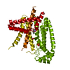
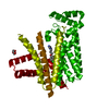
 Movie
Movie Controller


+ Open data

- Basic information
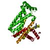
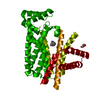
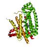
| Entry | Database: PDB / ID: 5qtj | ||||||
|---|---|---|---|---|---|---|---|
| Title | T. brucei FPPS in complex with CID 47256035 | ||||||
 Components Components | Farnesyl pyrophosphate synthase | ||||||
 Keywords Keywords | Transferase/Transferase inhibitor / farnesyl diphosphate synthase / Trypanosoma brucei / PanDDA / protein-ligand complex / Transferase-Transferase inhibitor complex | ||||||
| Function / homology |  Function and homology information Function and homology informationtrans, trans-farnesyl diphosphate biosynthetic process / dimethylallyltranstransferase activity / (2E,6E)-farnesyl diphosphate synthase activity / metal ion binding / cytoplasm Similarity search - Function | ||||||
| Biological species |  | ||||||
| Method |  X-RAY DIFFRACTION / SYNCHROTRON / MOLECULAR REPLACEMENT / molecular replacement / Resolution: 2.101 Å X-RAY DIFFRACTION / SYNCHROTRON / MOLECULAR REPLACEMENT / molecular replacement / Resolution: 2.101 Å | ||||||
 Authors Authors | Muenzker, L. / Petrick, J.K. / Schleberger, C. / Cornaciu, I. / Marquez, J.A. / Jahnke, W. | ||||||
 Citation Citation | Journal: To Be Published Title: T. brucei FPPS in complex with CID 47256035 Authors: Muenzker, L. | ||||||
| History |
|
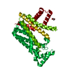
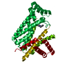
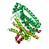
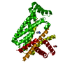
- Structure visualization
| Structure viewer | Molecule: MolmilJmol/JSmol |
|---|
- Downloads & links
-Download
| PDBx/mmCIF format | 5qtj.cif.gz | 153.5 KB | Display | PDBx/mmCIF format |
|---|---|---|---|---|
| PDB format | pdb5qtj.ent.gz | 120.9 KB | Display | PDB format |
| PDBx/mmJSON format | 5qtj.json.gz | Tree view | PDBx/mmJSON format | |
| Others |  Other downloads Other downloads |
-Validation report
| Arichive directory | https://data.pdbj.org/pub/pdb/validation_reports/qt/5qtjftp://data.pdbj.org/pub/pdb/validation_reports/qt/5qtj | HTTPS FTP |
|---|
-Group deposition
| ID | G_1002102 (7 entries) |
|---|---|
| Title | PanDDA analysis group deposition |
| Type | changed state |
| Description | FPPS screened against the Enamine Golden Fragment Library by X-ray Crystallography at the HTX lab of EMBL Grenoble |


-Related structure data
| Related structure data | 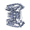 4rypS S: Starting model for refinement |
|---|---|
| Similar structure data |


-Links
 PDBj
PDBj
- Assembly
| Deposited unit | 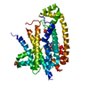
| |||||||||
|---|---|---|---|---|---|---|---|---|---|---|
| 1 | 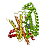
| |||||||||
| Unit cell |
| |||||||||
| Components on special symmetry positions |
|
-Components
-Protein , 1 types, 1 molecules A
| #1: Protein | Mass: 42169.211 Da / Num. of mol.: 1 Source method: isolated from a genetically manipulated source Source: (gene. exp.)  |
|---|
-Non-polymers , 5 types, 92 molecules 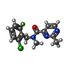


| #2: Chemical | ChemComp-PJS /  Mass: 293.724 Da / Num. of mol.: 1 / Source method: obtained synthetically / Formula: C14H13ClFN3O Mass: 293.724 Da / Num. of mol.: 1 / Source method: obtained synthetically / Formula: C14H13ClFN3O | ||||||
|---|---|---|---|---|---|---|---|
| #3: Chemical |  Mass: 94.971 Da / Num. of mol.: 2 / Source method: obtained synthetically / Formula: PO4 Mass: 94.971 Da / Num. of mol.: 2 / Source method: obtained synthetically / Formula: PO4#4: Chemical | ChemComp-NA / |  Mass: 22.990 Da / Num. of mol.: 1 / Source method: obtained synthetically / Formula: Na Mass: 22.990 Da / Num. of mol.: 1 / Source method: obtained synthetically / Formula: Na#5: Chemical | ChemComp-GOL / |  Mass: 92.094 Da / Num. of mol.: 1 / Source method: obtained synthetically / Formula: C3H8O3 Mass: 92.094 Da / Num. of mol.: 1 / Source method: obtained synthetically / Formula: C3H8O3#6: Water | ChemComp-HOH / | Mass: 18.015 Da / Num. of mol.: 87 / Source method: isolated from a natural source / Formula: H2O |
-Details
| Has protein modification | N |
|---|
-Experimental details
-Experiment
| Experiment | Method: X-RAY DIFFRACTION / Number of used crystals: 1 |
|---|
- Sample preparation
| Crystal | Density Matthews: 2.15 Å3/Da / Density % sol: 42.69 % / Mosaicity: 0.05 ° |
|---|---|
| Crystal grow | Temperature: 293 K / Method: vapor diffusion, sitting drop / pH: 6 / Details: 0.12 M CsCl 12 % w/v PEG 3350, 12 % v/v DMSO |
-Data collection
| Diffraction | Mean temperature: 100 K |
|---|---|
| Diffraction source | Source: SYNCHROTRON / Site: ESRF  / Beamline: MASSIF-1 / Wavelength: 0.966 Å / Beamline: MASSIF-1 / Wavelength: 0.966 Å |
| Detector | Type: DECTRIS PILATUS3 2M / Detector: PIXEL / Date: Jun 4, 2018 |
| Radiation | Protocol: SINGLE WAVELENGTH / Monochromatic (M) / Laue (L): M / Scattering type: x-ray |
| Radiation wavelength | Wavelength: 0.966 Å / Relative weight: 1 |
| Reflection | Resolution: 2.101→57.028 Å / Num. obs: 17890 / % possible obs: 77.9 % / Redundancy: 35.2 % / CC1/2: 0.999 / Rmerge(I) obs: 0.134 / Rpim(I) all: 0.021 / Rrim(I) all: 0.128 / Rsym value: 0.134 / Net I/σ(I): 21.7 / Num. measured all: 590391 / Scaling rejects: 2 |
| Reflection shell | Resolution: 2.101→2.282 Å / Redundancy: 20.7 % / Rmerge(I) obs: 1.927 / Mean I/σ(I) obs: 1.5 / Num. measured all: 83677 / Num. unique obs: 2286 / CC1/2: 0.952 / Rpim(I) all: 0.247 / Rrim(I) all: 1.504 / Rsym value: 1.927 / Net I/σ(I) obs: 2.7 / % possible all: 18.4 |
-Phasing
| Phasing | Method: molecular replacement |
|---|
- Processing
| Software |
| |||||||||||||||||||||||||||||||||||||||||||||||||
|---|---|---|---|---|---|---|---|---|---|---|---|---|---|---|---|---|---|---|---|---|---|---|---|---|---|---|---|---|---|---|---|---|---|---|---|---|---|---|---|---|---|---|---|---|---|---|---|---|---|---|
| Refinement | Method to determine structure: MOLECULAR REPLACEMENT Starting model: pdbid 4ryp Resolution: 2.101→57.028 Å / SU ML: 0.25 / Cross valid method: THROUGHOUT / σ(F): 1.34 / Phase error: 31.81 / Stereochemistry target values: ML
| |||||||||||||||||||||||||||||||||||||||||||||||||
| Solvent computation | Shrinkage radii: 0.9 Å / VDW probe radii: 1.11 Å / Solvent model: FLAT BULK SOLVENT MODEL | |||||||||||||||||||||||||||||||||||||||||||||||||
| Displacement parameters | Biso max: 154.76 Å2 / Biso mean: 56.8529 Å2 / Biso min: 17.44 Å2 | |||||||||||||||||||||||||||||||||||||||||||||||||
| Refinement step | Cycle: final / Resolution: 2.101→57.028 Å
| |||||||||||||||||||||||||||||||||||||||||||||||||
| LS refinement shell | Refine-ID: X-RAY DIFFRACTION / Rfactor Rfree error: 0 / Total num. of bins used: 6
|
