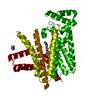
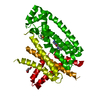
 Movie
Movie Controller


+ Open data

- Basic information
| Entry | Database: PDB / ID: 5qt6 | ||||||
|---|---|---|---|---|---|---|---|
| Title | T. brucei FPPS in complex with CID 112445 | ||||||
 Components Components | Farnesyl pyrophosphate synthase | ||||||
 Keywords Keywords | TRANSFERASE/TRANSFERASE inhibitor / farnesyl diphosphate synthase / Trypanosoma brucei / PanDDA / protein-ligand complex / TRANSFERASE-TRANSFERASE inhibitor complex | ||||||
| Function / homology |  Function and homology information Function and homology informationtrans, trans-farnesyl diphosphate biosynthetic process / dimethylallyltranstransferase activity / (2E,6E)-farnesyl diphosphate synthase activity / metal ion binding / cytoplasm Similarity search - Function | ||||||
| Biological species |  | ||||||
| Method |  X-RAY DIFFRACTION / SYNCHROTRON / MOLECULAR REPLACEMENT / molecular replacement / Resolution: 2.189 Å X-RAY DIFFRACTION / SYNCHROTRON / MOLECULAR REPLACEMENT / molecular replacement / Resolution: 2.189 Å | ||||||
 Authors Authors | Muenzker, L. / Petrick, J.K. / Schleberger, C. / Jahnke, W. | ||||||
 Citation Citation | Journal: To Be Published Title: T. brucei FPPS in complex with CID 112445 Authors: Muenzker, L. | ||||||
| History |
|
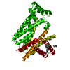
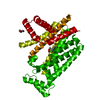
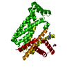
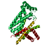
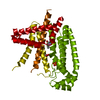
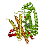
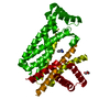
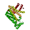
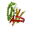
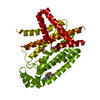
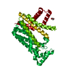
- Structure visualization
| Structure viewer | Molecule: MolmilJmol/JSmol |
|---|
- Downloads & links
-Download
| PDBx/mmCIF format | 5qt6.cif.gz | 148.8 KB | Display | PDBx/mmCIF format |
|---|---|---|---|---|
| PDB format | pdb5qt6.ent.gz | 117.6 KB | Display | PDB format |
| PDBx/mmJSON format | 5qt6.json.gz | Tree view | PDBx/mmJSON format | |
| Others |  Other downloads Other downloads |
-Validation report
| Arichive directory | https://data.pdbj.org/pub/pdb/validation_reports/qt/5qt6ftp://data.pdbj.org/pub/pdb/validation_reports/qt/5qt6 | HTTPS FTP |
|---|
-Group deposition
| ID | G_1002098 (8 entries) |
|---|---|
| Title | PanDDA analysis group deposition |
| Type | changed state |
| Description | X-ray crystallography of TbruFPPS at Novartis, Basel and data collection at SLS beamline X10SA |


-Related structure data
| Related structure data | 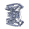 4rypS S: Starting model for refinement |
|---|---|
| Similar structure data |


-Links
 PDBj
PDBj
- Assembly
| Deposited unit | 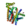
| ||||||||||||
|---|---|---|---|---|---|---|---|---|---|---|---|---|---|
| 1 | 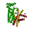
| ||||||||||||
| Unit cell |
| ||||||||||||
| Components on special symmetry positions |
|
-Components
| #1: Protein | Mass: 42169.211 Da / Num. of mol.: 1 Source method: isolated from a genetically manipulated source Source: (gene. exp.)  |
|---|---|
| #2: Chemical | ChemComp-JNE / 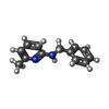  Mass: 198.264 Da / Num. of mol.: 1 / Source method: obtained synthetically / Formula: C13H14N2 / Feature type: SUBJECT OF INVESTIGATION Mass: 198.264 Da / Num. of mol.: 1 / Source method: obtained synthetically / Formula: C13H14N2 / Feature type: SUBJECT OF INVESTIGATION |
| #3: Chemical | ChemComp-PEG /   Mass: 106.120 Da / Num. of mol.: 1 / Source method: obtained synthetically / Formula: C4H10O3 Mass: 106.120 Da / Num. of mol.: 1 / Source method: obtained synthetically / Formula: C4H10O3 |
| #4: Chemical | ChemComp-DMS /   Mass: 78.133 Da / Num. of mol.: 1 / Source method: obtained synthetically / Formula: C2H6OS / Comment: DMSO, precipitant*YM Mass: 78.133 Da / Num. of mol.: 1 / Source method: obtained synthetically / Formula: C2H6OS / Comment: DMSO, precipitant*YM |
| #5: Water | ChemComp-HOH /  Mass: 18.015 Da / Num. of mol.: 66 / Source method: isolated from a natural source / Formula: H2O Mass: 18.015 Da / Num. of mol.: 66 / Source method: isolated from a natural source / Formula: H2O |
| Has ligand of interest | Y |
| Has protein modification | N |
-Experimental details
-Experiment
| Experiment | Method: X-RAY DIFFRACTION / Number of used crystals: 1 |
|---|
- Sample preparation
| Crystal | Density Matthews: 2.15 Å3/Da / Density % sol: 42.91 % / Mosaicity: 0.09 ° |
|---|---|
| Crystal grow | Temperature: 293 K / Method: vapor diffusion, sitting drop / pH: 6 / Details: 0.12 M CsCl 12 % w/v PEG 3350, 6 % v/v DMSO |
-Data collection
| Diffraction | Mean temperature: 100 K |
|---|---|
| Diffraction source | Source: SYNCHROTRON / Site: SLS  / Beamline: X10SA / Wavelength: 0.99979 Å / Beamline: X10SA / Wavelength: 0.99979 Å |
| Detector | Type: DECTRIS PILATUS3 6M / Detector: PIXEL / Date: May 26, 2017 |
| Radiation | Protocol: SINGLE WAVELENGTH / Monochromatic (M) / Laue (L): M / Scattering type: x-ray |
| Radiation wavelength | Wavelength: 0.99979 Å / Relative weight: 1 |
| Reflection | Resolution: 2.189→52.554 Å / Num. obs: 12355 / % possible obs: 60.3 % / Redundancy: 17.6 % / CC1/2: 0.998 / Rmerge(I) obs: 0.477 / Rpim(I) all: 0.233 / Rrim(I) all: 1.013 / Rsym value: 0.477 / Net I/σ(I): 11 / Num. measured all: 280753 / Scaling rejects: 461 |
| Reflection shell | Resolution: 2.189→2.429 Å / Redundancy: 18 % / Rmerge(I) obs: 2.049 / Mean I/σ(I) obs: 1.8 / Num. measured all: 42463 / Num. unique obs: 2206 / CC1/2: 0.443 / Rpim(I) all: 5.932 / Rrim(I) all: 26.373 / Rsym value: 2.049 / Net I/σ(I) obs: 0.9 / % possible all: 11.7 |
-Phasing
| Phasing | Method: molecular replacement |
|---|
- Processing
| Software |
| ||||||||||||||||||||||||||||||||||||||||||
|---|---|---|---|---|---|---|---|---|---|---|---|---|---|---|---|---|---|---|---|---|---|---|---|---|---|---|---|---|---|---|---|---|---|---|---|---|---|---|---|---|---|---|---|
| Refinement | Method to determine structure: MOLECULAR REPLACEMENT Starting model: pdbid 4ryp Resolution: 2.189→52.554 Å / SU ML: 0.25 / Cross valid method: THROUGHOUT / σ(F): 1.33 / Phase error: 30.35 / Stereochemistry target values: ML
| ||||||||||||||||||||||||||||||||||||||||||
| Solvent computation | Shrinkage radii: 0.9 Å / VDW probe radii: 1.11 Å / Solvent model: FLAT BULK SOLVENT MODEL | ||||||||||||||||||||||||||||||||||||||||||
| Displacement parameters | Biso max: 178.32 Å2 / Biso mean: 56.4189 Å2 / Biso min: 16.94 Å2 | ||||||||||||||||||||||||||||||||||||||||||
| Refinement step | Cycle: final / Resolution: 2.189→52.554 Å
| ||||||||||||||||||||||||||||||||||||||||||
| LS refinement shell | Refine-ID: X-RAY DIFFRACTION / Rfactor Rfree error: 0 / Total num. of bins used: 5
|
