[English] 日本語
Yorodumi
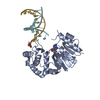
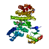
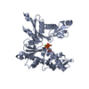
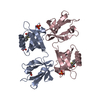
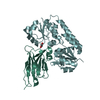
- PDB-5ovw: Nanobody-bound BtuF, the vitamin B12 binding protein in Escherich... -

+
Open data


ID or keywords:

Loading...

-
Basic information

Entry
Database: PDB / ID: 5ovw
TitleNanobody-bound BtuF, the vitamin B12 binding protein in Escherichia coli
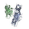
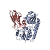
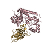
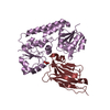
Components
  • NanobodySingle-domain antibody
  • Vitamin B12-binding protein
KeywordsTRANSPORT PROTEIN / Nanobody / Inhibitor / vitamin B12 / substrate-binding protein
Function / homology
Function and homology information


cobalamin transport complex / cobalamin transport / cobalamin binding / outer membrane-bounded periplasmic space / periplasmic space / membrane
Similarity search - Function
ABC transporter, vitamin B12-binding protein / ABC transporter periplasmic binding domain / Periplasmic binding protein / Iron siderophore/cobalamin periplasmic-binding domain profile. / Nitrogenase molybdenum iron protein domain / Rossmann fold / 3-Layer(aba) Sandwich / Alpha Beta
Similarity search - Domain/homology
Vitamin B12-binding protein
Similarity search - Component
Biological speciesEscherichia coli (E. coli)
Lama glama (llama)
MethodX-RAY DIFFRACTION / SYNCHROTRON / MOLECULAR REPLACEMENT / Resolution: 2.653 Å
AuthorsMireku, S.A. / Sauer, M.M. / Glockshuber, R. / Locher, K.P.
Funding support Switzerland, 1items
OrganizationGrant numberCountry
Swiss National Science FoundationSNF 310030B_166672 Switzerland
CitationJournal: Sci Rep / Year: 2017
Title: Structural basis of nanobody-mediated blocking of BtuF, the cognate substrate-binding protein of the Escherichia coli vitamin B12 transporter BtuCD.
Authors: Mireku, S.A. / Sauer, M.M. / Glockshuber, R. / Locher, K.P.
History
DepositionAug 30, 2017Deposition site: PDBE / Processing site: PDBE
Revision 1.0Nov 8, 2017Provider: repository / Type: Initial release
Revision 1.1Oct 16, 2019Group: Data collection / Category: reflns_shell

-
Structure visualization

Structure viewerMolecule:
MolmilJmol/JSmol

Downloads & links

-
Assembly

Deposited unit
A: Vitamin B12-binding protein
B: Vitamin B12-binding protein
C: Vitamin B12-binding protein
D: Vitamin B12-binding protein
E: Vitamin B12-binding protein
F: Vitamin B12-binding protein
G: Nanobody
H: Nanobody
I: Nanobody
J: Nanobody
K: Nanobody
L: Nanobody
hetero molecules


Theoretical massNumber of molelcules
Total (without water)291,32217
Polymers290,86112
Non-polymers4605
Water4,756264
1
A: Vitamin B12-binding protein
H: Nanobody
hetero molecules


Theoretical massNumber of molelcules
Total (without water)48,5693
Polymers48,4772
Non-polymers921
Water362
TypeNameSymmetry operationNumber
identity operation1_555x,y,z1
Buried area2470 Å2
ΔGint-14 kcal/mol
Surface area16110 Å2
MethodPISA
2
B: Vitamin B12-binding protein
I: Nanobody
hetero molecules


Theoretical massNumber of molelcules
Total (without water)48,5693
Polymers48,4772
Non-polymers921
Water362
TypeNameSymmetry operationNumber
identity operation1_555x,y,z1
Buried area2450 Å2
ΔGint-13 kcal/mol
Surface area16220 Å2
MethodPISA
3
C: Vitamin B12-binding protein
G: Nanobody


Theoretical massNumber of molelcules
Total (without water)48,4772
Polymers48,4772
Non-polymers00
Water362
TypeNameSymmetry operationNumber
identity operation1_555x,y,z1
Buried area2160 Å2
ΔGint-12 kcal/mol
Surface area16310 Å2
MethodPISA
4
D: Vitamin B12-binding protein
J: Nanobody
hetero molecules


Theoretical massNumber of molelcules
Total (without water)48,5693
Polymers48,4772
Non-polymers921
Water362
TypeNameSymmetry operationNumber
identity operation1_555x,y,z1
Buried area2480 Å2
ΔGint-14 kcal/mol
Surface area16200 Å2
MethodPISA
5
E: Vitamin B12-binding protein
K: Nanobody
hetero molecules


Theoretical massNumber of molelcules
Total (without water)48,5693
Polymers48,4772
Non-polymers921
Water362
TypeNameSymmetry operationNumber
identity operation1_555x,y,z1
Buried area2480 Å2
ΔGint-13 kcal/mol
Surface area16260 Å2
MethodPISA
6
F: Vitamin B12-binding protein
L: Nanobody
hetero molecules


Theoretical massNumber of molelcules
Total (without water)48,5693
Polymers48,4772
Non-polymers921
Water362
TypeNameSymmetry operationNumber
identity operation1_555x,y,z1
Buried area2610 Å2
ΔGint-12 kcal/mol
Surface area16200 Å2
MethodPISA
Unit cell
Length a, b, c (Å)83.800, 141.000, 216.700
Angle α, β, γ (deg.)90.00, 90.00, 90.00
Int Tables number19
Space group name H-MP212121

-
Components

#1: Protein
Vitamin B12-binding protein


Mass: 31511.924 Da / Num. of mol.: 6
Source method: isolated from a genetically manipulated source
Details: OmpA-BtuF-3C-His6 / Source: (gene. exp.) Escherichia coli (E. coli) / Gene: btuF, yadT, b0158, JW0154 / Production host: Escherichia coli (E. coli) / References: UniProt: P37028
#2: Antibody
Nanobody / Single-domain antibody


Mass: 16964.984 Da / Num. of mol.: 6
Source method: isolated from a genetically manipulated source
Details: SecretionSignal-Nanobody-His6 / Source: (gene. exp.) Lama glama (llama) / Production host: Escherichia coli (E. coli)
#3: Chemical
ChemComp-GOL / GLYCEROL / GLYCERIN / PROPANE-1,2,3-TRIOL / Glycerol


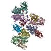
Mass: 92.094 Da / Num. of mol.: 5 / Source method: obtained synthetically / Formula: C3H8O3
#4: Water ChemComp-HOH / water / Water


Mass: 18.015 Da / Num. of mol.: 264 / Source method: isolated from a natural source / Formula: H2O

-
Experimental details

-
Experiment

ExperimentMethod: X-RAY DIFFRACTION / Number of used crystals: 1

-
Sample preparation

CrystalDensity Matthews: 2.2 Å3/Da / Density % sol: 44.11 %
Crystal growTemperature: 293.15 K / Method: vapor diffusion, sitting drop / Details: PEG4000, Tris-HCl, Magnesium chloride

-
Data collection

DiffractionMean temperature: 100 K
Diffraction sourceSource: SYNCHROTRON / Site: SLS / Beamline: X06SA / Wavelength: 0.99984 Å
DetectorType: DECTRIS PILATUS 6M-F / Detector: PIXEL / Date: Sep 30, 2016
RadiationProtocol: SINGLE WAVELENGTH / Monochromatic (M) / Laue (L): M / Scattering type: x-ray
Radiation wavelengthWavelength: 0.99984 Å / Relative weight: 1
ReflectionResolution: 2.653→19.996 Å / Num. obs: 63910 / % possible obs: 85.33 % / Redundancy: 6.8 % / Net I/σ(I): 1.36

-
Processing

Software
NameVersionClassification
PHENIX(1.11.1_2575: ???)refinement
Cootmodel building
XDSdata scaling
PHENIXphasing
RefinementMethod to determine structure: MOLECULAR REPLACEMENT / Resolution: 2.653→19.996 Å / SU ML: 0.32 / Cross valid method: FREE R-VALUE / σ(F): 1.36 / Phase error: 28.99 / Stereochemistry target values: ML
RfactorNum. reflection% reflection
Rfree0.2594 3239 5.07 %
Rwork0.2232 --
obs0.2251 63910 85.33 %
Solvent computationShrinkage radii: 0.9 Å / VDW probe radii: 1.11 Å / Solvent model: FLAT BULK SOLVENT MODEL
Refinement stepCycle: LAST / Resolution: 2.653→19.996 Å
ProteinNucleic acidLigandSolventTotal
Num. atoms17058 0 30 264 17352
Refine LS restraints
Refine-IDTypeDev idealNumber
X-RAY DIFFRACTIONf_bond_d0.00317479
X-RAY DIFFRACTIONf_angle_d0.55823797
X-RAY DIFFRACTIONf_dihedral_angle_d12.98710594
X-RAY DIFFRACTIONf_chiral_restr0.0432622
X-RAY DIFFRACTIONf_plane_restr0.0043126
LS refinement shell
Resolution (Å)Rfactor RfreeNum. reflection RfreeRfactor RworkNum. reflection RworkRefine-ID% reflection obs (%)
2.6533-2.69280.505480.382174X-RAY DIFFRACTION6
2.6928-2.73480.3261290.3829508X-RAY DIFFRACTION17
2.7348-2.77950.4048480.3627927X-RAY DIFFRACTION30
2.7795-2.82730.33871010.34821534X-RAY DIFFRACTION51
2.8273-2.87860.42761280.3412176X-RAY DIFFRACTION71
2.8786-2.93380.35481330.34012632X-RAY DIFFRACTION87
2.9338-2.99350.35131520.33233030X-RAY DIFFRACTION98
2.9935-3.05830.33561720.33523026X-RAY DIFFRACTION100
3.0583-3.12920.34531750.32173059X-RAY DIFFRACTION100
3.1292-3.20710.34211420.29953085X-RAY DIFFRACTION100
3.2071-3.29350.29491500.29953066X-RAY DIFFRACTION100
3.2935-3.390.34511460.29023101X-RAY DIFFRACTION100
3.39-3.49880.29581500.26633115X-RAY DIFFRACTION100
3.4988-3.62320.28591580.24523076X-RAY DIFFRACTION100
3.6232-3.76730.30851740.24453051X-RAY DIFFRACTION100
3.7673-3.93750.28581800.2173074X-RAY DIFFRACTION100
3.9375-4.14340.22841610.19173097X-RAY DIFFRACTION100
4.1434-4.40040.2121670.17273124X-RAY DIFFRACTION100
4.4004-4.7360.21751660.16313091X-RAY DIFFRACTION100
4.736-5.20490.20151750.16213118X-RAY DIFFRACTION100
5.2049-5.94080.21131700.17223146X-RAY DIFFRACTION100
5.9408-7.42060.21371630.18223190X-RAY DIFFRACTION100
7.4206-19.99690.16981910.15043271X-RAY DIFFRACTION100
Refinement TLS params.

Method: refined / Refine-ID: X-RAY DIFFRACTION

IDL112)L122)L132)L222)L232)L332)S11 (Å °)S12 (Å °)S13 (Å °)S21 (Å °)S22 (Å °)S23 (Å °)S31 (Å °)S32 (Å °)S33 (Å °)T112)T122)T132)T222)T232)T332)Origin x (Å)Origin y (Å)Origin z (Å)
10.17080.08530.07570.14710.00040.24540.0865-0.06530.03450.16990.09020.2041-0.0247-0.10830.07220.43550.00530.08990.3995-0.02020.52184.9604153.38236.6091
20.107-0.1471-0.03770.1897-0.02420.0560.08350.0350.11660.1325-0.0554-0.08910.0285-0.054400.3814-0.055-0.0340.4025-0.04940.514231.5942149.78255.6018
30.65380.11490.02210.27960.11270.5772-0.36030.13520.2691-0.19530.13960.2694-0.37220.0086-0.41690.6229-0.0854-0.15660.44890.02970.45512.0563122.2943-29.013
40.12080.02840.04130.7057-0.11140.0564-0.05490.2092-0.2876-0.01080.33910.04540.12650.440.47940.31830.1657-0.01180.6449-0.18680.39911.90197.2695-27.5641
50.1713-0.08670.07730.23030.22240.2020.1489-0.15220.09180.1574-0.01530.20370.0062-0.05640.26760.5495-0.03050.19090.486-0.13550.51354.9192157.375728.3533
60.03590.02770.00770.12120.10120.07830.1510.1570.55110.02940.1346-0.1939-0.29220.11910.0230.7671-0.25990.3530.8188-0.10610.943816.2098181.824526.5416
70.16250.07220.22040.1579-0.03340.25030.0007-0.14550.25620.0161-0.12750.2706-0.1105-0.0711-0.18360.36760.0103-0.06350.4194-0.11510.50753.5659125.9294-7.2848
80.12490.1520.07860.2210.04390.0756-0.075-0.02090.1508-0.11960.0603-0.0424-0.14610.0437-0.00070.2941-0.04030.03720.3676-0.06080.420330.4807127.8683-7.731
90.12840.26710.13240.50150.21190.3321-0.0520.096-0.20580.1720.1824-0.01160.2841-0.11540.2420.3834-0.04740.09340.48280.05320.31621.0245101.499943.9537
100.2373-0.12180.02390.2622-0.14780.1497-0.2302-0.24880.05130.49930.2994-0.1043-0.17560.08370.12230.63190.0294-0.06590.4191-0.12020.374635.3883124.423942.2568
110.44020.3465-0.1230.51810.28680.38930.2160.29940.05530.30920.0087-0.06160.2315-0.12310.08470.7213-0.07840.11090.41680.02060.415118.3527102.289665.7283
120.00610.00930.02820.03750.03970.0821-0.45880.06920.2084-0.2940.0720.5410.1755-0.1697-0.15670.85860.0905-0.01140.56020.24550.94290.0944122.088165.1581
130.2391-0.1479-0.06150.1048-0.00270.05080.2941-0.02050.17340.4209-0.3484-0.4244-0.08610.1503-0.00740.824-0.2305-0.10380.9643-0.33290.902731.75160.545741.9097
140.10550.1370.01560.2177-0.10790.1301-0.11890.04430.0748-0.17510.2037-0.16670.0074-0.12290.08480.5429-0.09390.06250.28640.00390.4419.7757172.9447-10.9603
150.419-0.2024-0.51110.14530.13080.6675-0.31760.52490.05520.0189-0.2754-0.065-0.06480.7385-1.09110.3069-0.52190.37682.0021-0.0655-0.0727.7344118.0333-43.8302
160.0899-0.018-0.03280.26830.31870.38480.1422-0.0556-0.2093-0.0730.0095-0.06130.0327-0.01850.23160.2471-0.0323-0.05350.4121-0.01220.389918.0617105.27688.9181
170.0675-0.1175-0.04110.33130.11240.0319-0.1497-0.1916-0.15560.33990.28710.2818-0.0755-0.38030.16090.38860.08490.12010.67590.14810.44038.38123.389627.8456
180.04850.09520.10280.08940.08260.1191-0.0643-0.00580.1127-0.20360.13520.0883-0.0550.10460.10540.8211-0.2830.19840.4981-0.1680.644426.4387127.561479.6433
Refinement TLS group
IDRefine-IDRefine TLS-IDSelection details
1X-RAY DIFFRACTION1chain 'A' and (resid 22 through 149 )
2X-RAY DIFFRACTION2chain 'A' and (resid 150 through 266 )
3X-RAY DIFFRACTION3chain 'B' and (resid 22 through 149 )
4X-RAY DIFFRACTION4chain 'B' and (resid 150 through 266 )
5X-RAY DIFFRACTION5chain 'C' and (resid 22 through 149 )
6X-RAY DIFFRACTION6chain 'C' and (resid 150 through 266 )
7X-RAY DIFFRACTION7chain 'D' and (resid 22 through 149 )
8X-RAY DIFFRACTION8chain 'D' and (resid 150 through 266 )
9X-RAY DIFFRACTION9chain 'E' and (resid 22 through 149 )
10X-RAY DIFFRACTION10chain 'E' and (resid 150 through 266 )
11X-RAY DIFFRACTION11chain 'F' and (resid 22 through 149 )
12X-RAY DIFFRACTION12chain 'F' and (resid 150 through 266 )
13X-RAY DIFFRACTION13chain 'G' and (resid 24 through 149 )
14X-RAY DIFFRACTION14chain 'H' and (resid 24 through 149 )
15X-RAY DIFFRACTION15chain 'I' and (resid 24 through 149 )
16X-RAY DIFFRACTION16chain 'J' and (resid 24 through 149 )
17X-RAY DIFFRACTION17chain 'K' and (resid 24 through 149 )
18X-RAY DIFFRACTION18chain 'L' and (resid 24 through 149 )

+
About Yorodumi

-
News

-
Feb 9, 2022. New format data for meta-information of EMDB entries

New format data for meta-information of EMDB entries

  • Version 3 of the EMDB header file is now the official format.
  • The previous official version 1.9 will be removed from the archive.

Related info.:EMDB header

External links:wwPDB to switch to version 3 of the EMDB data model

-
Aug 12, 2020. Covid-19 info

Covid-19 info

URL: https://pdbj.org/emnavi/covid19.php

New page: Covid-19 featured information page in EM Navigator.

Related info.:Covid-19 info / Mar 5, 2020. Novel coronavirus structure data

+
Mar 5, 2020. Novel coronavirus structure data

Novel coronavirus structure data

Related info.:Yorodumi Speices / Aug 12, 2020. Covid-19 info

External links:COVID-19 featured content - PDBj / Molecule of the Month (242):Coronavirus Proteases

+
Jan 31, 2019. EMDB accession codes are about to change! (news from PDBe EMDB page)

EMDB accession codes are about to change! (news from PDBe EMDB page)

  • The allocation of 4 digits for EMDB accession codes will soon come to an end. Whilst these codes will remain in use, new EMDB accession codes will include an additional digit and will expand incrementally as the available range of codes is exhausted. The current 4-digit format prefixed with “EMD-” (i.e. EMD-XXXX) will advance to a 5-digit format (i.e. EMD-XXXXX), and so on. It is currently estimated that the 4-digit codes will be depleted around Spring 2019, at which point the 5-digit format will come into force.
  • The EM Navigator/Yorodumi systems omit the EMD- prefix.

Related info.:Q: What is EMD? / ID/Accession-code notation in Yorodumi/EM Navigator

External links:EMDB Accession Codes are Changing Soon! / Contact to PDBj

+
Jul 12, 2017. Major update of PDB

Major update of PDB

  • wwPDB released updated PDB data conforming to the new PDBx/mmCIF dictionary.
  • This is a major update changing the version number from 4 to 5, and with Remediation, in which all the entries are updated.
  • In this update, many items about electron microscopy experimental information are reorganized (e.g. em_software).
  • Now, EM Navigator and Yorodumi are based on the updated data.

External links:wwPDB Remediation / Enriched Model Files Conforming to OneDep Data Standards Now Available in the PDB FTP Archive

-
Yorodumi

Thousand views of thousand structures

  • Yorodumi is a browser for structure data from EMDB, PDB, SASBDB, etc.
  • This page is also the successor to EM Navigator detail page, and also detail information page/front-end page for Omokage search.
  • The word "yorodu" (or yorozu) is an old Japanese word meaning "ten thousand". "mi" (miru) is to see.

Related info.:EMDB / PDB / SASBDB / Comparison of 3 databanks / Yorodumi Search / Aug 31, 2016. New EM Navigator & Yorodumi / Yorodumi Papers / Jmol/JSmol / Function and homology information / Changes in new EM Navigator and Yorodumi

Read more