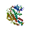
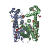
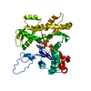
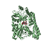
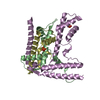
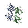
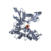
Journal: Proc Natl Acad Sci U S A / Year: 2016 Title: X-ray and cryo-EM structures of monomeric and filamentous actin-like protein MamK reveal changes associated with polymerization. Authors: Jan Löwe / Shaoda He / Sjors H W Scheres / Christos G Savva / Abstract: Magnetotactic bacteria produce iron-rich magnetic nanoparticles that are enclosed by membrane invaginations to form magnetosomes so they are able to sense and act upon Earth's magnetic field. In ...Magnetotactic bacteria produce iron-rich magnetic nanoparticles that are enclosed by membrane invaginations to form magnetosomes so they are able to sense and act upon Earth's magnetic field. In Magnetospirillum and other magnetotactic bacteria, to combine their magnetic moments, magnetosomes align along filaments formed by a bacterial actin homolog, MamK. Here, we present the crystal structure of a nonpolymerizing mutant of MamK from Magnetospirillum magneticum AMB-1 at 1.8-Å resolution, revealing its close similarity to actin and MreB. The crystals contain AMPPNP-bound monomeric MamK in two different conformations. To investigate conformational changes associated with polymerization, we used unmodified MamK protein and cryo-EM with helical 3D reconstruction in RELION to obtain a density map and a fully refined atomic model of MamK in filamentous form at 3.6-Å resolution. The filament is parallel (polar) double-helical, with a rise of 52.2 Å and a twist of 23.8°. As shown previously and unusually for actin-like filaments, the MamK subunits from each of the two strands are juxtaposed, creating an additional twofold axis along the filament. Compared with monomeric MamK, ADP-bound MamK in the filament undergoes a conformational change, rotating domains I and II against each other to further close the interdomain cleft between subdomains IB and IIB. The domain movement causes several loops to close around the nucleotide-binding pocket. Glu-143, a key residue for catalysis coordinating the magnesium ion, moves closer, presumably switching nucleotide hydrolysis upon polymerization-one of the hallmarks of cytomotive filaments of the actin type.
In the structure databanks used in Yorodumi, some data are registered as the other names, "COVID-19 virus" and "2019-nCoV". Here are the details of the virus and the list of structure data.
Jan 31, 2019. EMDB accession codes are about to change! (news from PDBe EMDB page)
EMDB accession codes are about to change! (news from PDBe EMDB page)
The allocation of 4 digits for EMDB accession codes will soon come to an end. Whilst these codes will remain in use, new EMDB accession codes will include an additional digit and will expand incrementally as the available range of codes is exhausted. The current 4-digit format prefixed with “EMD-” (i.e. EMD-XXXX) will advance to a 5-digit format (i.e. EMD-XXXXX), and so on. It is currently estimated that the 4-digit codes will be depleted around Spring 2019, at which point the 5-digit format will come into force.
The EM Navigator/Yorodumi systems omit the EMD- prefix.
Related info.:Q: What is EMD? / ID/Accession-code notation in Yorodumi/EM Navigator
Yorodumi is a browser for structure data from EMDB, PDB, SASBDB, etc.
This page is also the successor to EM Navigator detail page, and also detail information page/front-end page for Omokage search.
The word "yorodu" (or yorozu) is an old Japanese word meaning "ten thousand". "mi" (miru) is to see.
Related info.:EMDB / PDB / SASBDB / Comparison of 3 databanks / Yorodumi Search / Aug 31, 2016. New EM Navigator & Yorodumi / Yorodumi Papers / Jmol/JSmol / Function and homology information / Changes in new EM Navigator and Yorodumi
 Movie
Movie Controller


 Open data
 Basic information
 Components
 Keywords
 Function and homology information
 Magnetospirillum magneticum AMB-1 (bacteria)
 X-RAY DIFFRACTION /
  Authors
 United Kingdom, 2items
  Citation
 Structure visualization
 Downloads & links
 Other downloads
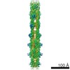


 PDBj
PDBj


 Assembly


 Mass: 24.305 Da / Num. of mol.: 2 / Source method: obtained synthetically / Formula: Mg
 Mass: 506.196 Da / Num. of mol.: 2 / Source method: obtained synthetically / Formula: C10H17N6O12P3 / Comment: AMP-PNP, energy-carrying molecule analogue*YM
 Mass: 18.015 Da / Num. of mol.: 536 / Source method: isolated from a natural source / Formula: H2O
 Sample preparation
 Processing