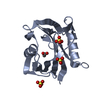
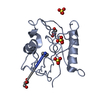
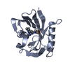
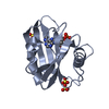
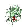
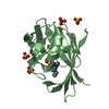
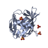
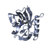
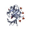
Entry Database : PDB / ID : 5otmTitle Crystal structure of human MTH1 in complex with O6-methyl-dGMP 7,8-dihydro-8-oxoguanine triphosphatase Keywords / / / / / / Function / homology Function Domain/homology Component
/ / / / / / / / / / / / / / / / / / / / / / / / / / / / / / / / / / / / / / / / / / / / / / / / / / / Biological species Homo sapiens (human)Method / / / Resolution : 1.8 Å Authors Gustafsson, R. / Henriksson, L. / Jemth, A.-S. / Brautigam, L. / Carreras Puigvert, J. / Homan, E. / Warpman Berglund, U. / Helleday, T. / Stenmark, P. Funding support Organization Grant number Country Swedish Research Council Knut and Alice Wallenberg Foundation Wenner-Gren Foundation Ake Wiberg Foundation Swedish Foundation for Strategic Research Swedish Pain Relief Foundation Torsten and Ragnar Soderberg Foundation Swedish Childrens Cancer Foundation Swedish Cancer Society
Journal : Nucleic Acids Res. / Year : 2018Title : MutT homologue 1 (MTH1) catalyzes the hydrolysis of mutagenic O6-methyl-dGTP.Authors : Jemth, A.S. / Gustafsson, R. / Brautigam, L. / Henriksson, L. / Vallin, K.S.A. / Sarno, A. / Almlof, I. / Homan, E. / Rasti, A. / Warpman Berglund, U. / Stenmark, P. / Helleday, T. History Deposition Aug 22, 2017 Deposition site / Processing site Revision 1.0 Sep 5, 2018 Provider / Type Revision 1.1 Oct 17, 2018 Group / Database references / Category Item _citation.country / _citation.journal_abbrev ... _citation.country / _citation.journal_abbrev / _citation.journal_id_ASTM / _citation.journal_id_CSD / _citation.journal_id_ISSN / _citation.pdbx_database_id_DOI / _citation.title / _citation.year Revision 1.2 Oct 24, 2018 Group / Database references / Category / citation_author / Item / _citation.titleRevision 1.3 Nov 28, 2018 Group / Database references / Category / pdbx_database_procItem / _citation.page_first / _citation.page_lastRevision 1.4 Jan 17, 2024 Group / Database references / Refinement descriptionCategory chem_comp_atom / chem_comp_bond ... chem_comp_atom / chem_comp_bond / database_2 / pdbx_initial_refinement_model Item / _database_2.pdbx_database_accession
Show all Show less
 Movie
Movie Controller


 Open data
 Basic information
 Components
 Keywords
 Function and homology information
 Homo sapiens (human)
 X-RAY DIFFRACTION /
  Authors
 Sweden, 9items
  Citation
 Structure visualization
 Downloads & links
 Other downloads
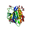


 PDBj
PDBj

 Assembly


 Mass: 96.063 Da / Num. of mol.: 8 / Source method: obtained synthetically / Formula: SO4
 Mass: 59.044 Da / Num. of mol.: 1 / Source method: obtained synthetically / Formula: C2H3O2
 Type: DNA linking / Mass: 361.248 Da / Num. of mol.: 2 / Source method: obtained synthetically / Formula: C11H16N5O7P / Feature type: SUBJECT OF INVESTIGATION
 Mass: 18.015 Da / Num. of mol.: 242 / Source method: isolated from a natural source / Formula: H2O
 Sample preparation
 / Beamline: 14.1 / Wavelength: 0.91801 Å
 Processing