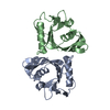
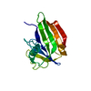
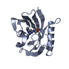
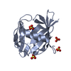
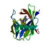
Entry Database : PDB / ID : 3zr1Title Crystal structure of human MTH1 7,8-DIHYDRO-8-OXOGUANINE TRIPHOSPHATASE Keywords Function / homology Function Domain/homology Component
/ / / / / / / / / / / / / / / / / / / / / / / / / / / / / / / / / / / / / / / / / / / / / / / / / / / Biological species HOMO SAPIENS (human)Method / / / Resolution : 1.9 Å Authors Svensson, L.M. / Jemth, A. / Desroses, M. / Loseva, O. / Helleday, T. / Hogbom, M. / Stenmark, P. Journal : FEBS Lett. / Year : 2011Title : Crystal Structure of Human Mth1 and the 8-Oxo-Dgmp Product Complex.Authors : Svensson, L.M. / Jemth, A. / Desroses, M. / Loseva, O. / Helleday, T. / Hogbom, M. / Stenmark, P. History Deposition Jun 13, 2011 Deposition site / Processing site Revision 1.0 Jul 27, 2011 Provider / Type Revision 1.1 Aug 24, 2011 Group Revision 1.2 Dec 20, 2023 Group Data collection / Database references ... Data collection / Database references / Derived calculations / Other / Refinement description Category chem_comp_atom / chem_comp_bond ... chem_comp_atom / chem_comp_bond / database_2 / pdbx_database_status / pdbx_initial_refinement_model / struct_site Item _database_2.pdbx_DOI / _database_2.pdbx_database_accession ... _database_2.pdbx_DOI / _database_2.pdbx_database_accession / _pdbx_database_status.status_code_sf / _struct_site.pdbx_auth_asym_id / _struct_site.pdbx_auth_comp_id / _struct_site.pdbx_auth_seq_id
Show all Show less
 Movie
Movie Controller


 Open data
 Basic information
 Components
 Keywords
 Function and homology information
 HOMO SAPIENS (human)
 X-RAY DIFFRACTION /
  Authors
 Citation
 Structure visualization
 Downloads & links
 Other downloads
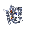


 PDBj
PDBj

 Assembly


 Mass: 59.044 Da / Num. of mol.: 2 / Source method: obtained synthetically / Formula: C2H3O2
 Mass: 96.063 Da / Num. of mol.: 4 / Source method: obtained synthetically / Formula: SO4
 Mass: 18.015 Da / Num. of mol.: 176 / Source method: isolated from a natural source / Formula: H2O
 Sample preparation
 / Beamline: 14.1 / Wavelength: 0.918
  Processing