 Movie
Movie Controller


+ Open data

- Basic information
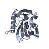
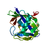
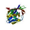
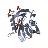
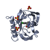
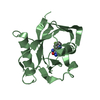
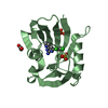
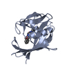
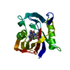
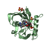
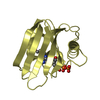
| Entry | Database: PDB / ID: 6jvo | ||||||
|---|---|---|---|---|---|---|---|
| Title | Crystal structure of human MTH1 in complex with compound MI1022 | ||||||
 Components Components | 7,8-dihydro-8-oxoguanine triphosphatase | ||||||
 Keywords Keywords | HYDROLASE / MTH1 / Oxidative DNA damage / 8-oxo-dGTP / Inhibitor development | ||||||
| Function / homology |  Function and homology information Function and homology information2-hydroxy-ATP hydrolase activity / 2-hydroxy-dATP hydrolase activity / N6-methyl-(d)ATP hydrolase activity / O6-methyl-dGTP hydrolase activity / 2-hydroxy-dATP diphosphatase / dATP diphosphatase activity / ATP diphosphatase activity / 8-oxo-7,8-dihydrodeoxyguanosine triphosphate pyrophosphatase activity / 8-oxo-7,8-dihydroguanosine triphosphate pyrophosphatase activity / Phosphate bond hydrolysis by NUDT proteins ...2-hydroxy-ATP hydrolase activity / 2-hydroxy-dATP hydrolase activity / N6-methyl-(d)ATP hydrolase activity / O6-methyl-dGTP hydrolase activity / 2-hydroxy-dATP diphosphatase / dATP diphosphatase activity / ATP diphosphatase activity / 8-oxo-7,8-dihydrodeoxyguanosine triphosphate pyrophosphatase activity / 8-oxo-7,8-dihydroguanosine triphosphate pyrophosphatase activity / Phosphate bond hydrolysis by NUDT proteins / DNA protection / hydrolase activity, acting on acid anhydrides, in phosphorus-containing anhydrides / purine nucleoside catabolic process / snoRNA binding / Hydrolases; Acting on acid anhydrides; In phosphorus-containing anhydrides / response to oxidative stress / mitochondrial matrix / DNA repair / mitochondrion / metal ion binding / nucleus / cytosol / cytoplasm Similarity search - Function | ||||||
| Biological species |  Homo sapiens (human) Homo sapiens (human) | ||||||
| Method |  X-RAY DIFFRACTION / SYNCHROTRON / MOLECULAR REPLACEMENT / Resolution: 1.902 Å X-RAY DIFFRACTION / SYNCHROTRON / MOLECULAR REPLACEMENT / Resolution: 1.902 Å | ||||||
 Authors Authors | Peng, C. / Li, Y.H. / Cheng, Y.S. | ||||||
 Citation Citation | Journal: Bioorg.Chem. / Year: 2021 Title: Inhibitor development of MTH1 via high-throughput screening with fragment based library and MTH1 substrate binding cavity. Authors: Peng, C. / Li, Y.H. / Yu, C.W. / Cheng, Z.H. / Liu, J.R. / Hsu, J.L. / Hsin, L.W. / Huang, C.T. / Juan, H.F. / Chern, J.W. / Cheng, Y.S. | ||||||
| History |
|
- Structure visualization
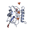
| Structure viewer | Molecule: MolmilJmol/JSmol |
|---|
- Downloads & links
-Download
| PDBx/mmCIF format | 6jvo.cif.gz | 150.1 KB | Display | PDBx/mmCIF format |
|---|---|---|---|---|
| PDB format | pdb6jvo.ent.gz | 116.1 KB | Display | PDB format |
| PDBx/mmJSON format | 6jvo.json.gz | Tree view | PDBx/mmJSON format | |
| Others |  Other downloads Other downloads |
-Validation report
| Arichive directory | https://data.pdbj.org/pub/pdb/validation_reports/jv/6jvoftp://data.pdbj.org/pub/pdb/validation_reports/jv/6jvo | HTTPS FTP |
|---|
-Related structure data
| Related structure data |  6jvfC  6jvgC  6jvhC 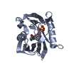 6jviC 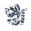 6jvjC 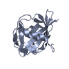 6jvkC 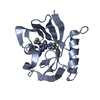 6jvlC 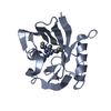 6jvmC 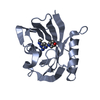 6jvnC 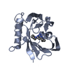 6jvpC 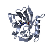 6jvqC 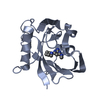 6jvrC 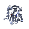 6jvsC 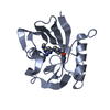 6jvtC 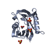 3zr1S S: Starting model for refinement C: citing same article ( |
|---|---|
| Similar structure data |


-Links
 PDBj
PDBj

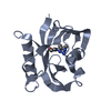
- Assembly
| Deposited unit | 
| |||||||||||||||||||||||||||||||||||||||||||||||||||||||||||||||||||||||
|---|---|---|---|---|---|---|---|---|---|---|---|---|---|---|---|---|---|---|---|---|---|---|---|---|---|---|---|---|---|---|---|---|---|---|---|---|---|---|---|---|---|---|---|---|---|---|---|---|---|---|---|---|---|---|---|---|---|---|---|---|---|---|---|---|---|---|---|---|---|---|---|---|
| 1 | 
| |||||||||||||||||||||||||||||||||||||||||||||||||||||||||||||||||||||||
| 2 | 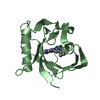
| |||||||||||||||||||||||||||||||||||||||||||||||||||||||||||||||||||||||
| Unit cell |
| |||||||||||||||||||||||||||||||||||||||||||||||||||||||||||||||||||||||
| Noncrystallographic symmetry (NCS) | NCS domain:
NCS domain segments: Ens-ID: 1
|