 Movie
Movie Controller

[English] 日本語
 Yorodumi
- PDB-5opk: Crystal structure of D52N/R367Q cN-II mutant bound to dATP and fr... -
+ Open data

- Basic information
| Entry | Database: PDB / ID: 5opk | ||||||
|---|---|---|---|---|---|---|---|
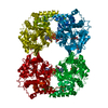
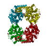
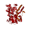
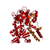
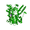
| Title | Crystal structure of D52N/R367Q cN-II mutant bound to dATP and free phosphate | ||||||
 Components Components | Cytosolic purine 5'-nucleotidase | ||||||
 Keywords Keywords | HYDROLASE / nucleotidase / relapsed leukemia | ||||||
| Function / homology |  Function and homology information Function and homology informationnucleoside phosphotransferase / : / nucleoside phosphotransferase activity / GMP metabolic process / Abacavir metabolism / dGMP metabolic process / negative regulation of defense response to virus by host / : / IMP-specific 5'-nucleotidase / dGMP catabolic process ...nucleoside phosphotransferase / : / nucleoside phosphotransferase activity / GMP metabolic process / Abacavir metabolism / dGMP metabolic process / negative regulation of defense response to virus by host / : / IMP-specific 5'-nucleotidase / dGMP catabolic process / adenosine metabolic process / Ribavirin ADME / IMP catabolic process / IMP metabolic process / allantoin metabolic process / Purine catabolism / 5'-nucleotidase / 5'-nucleotidase activity / protein K48-linked ubiquitination / ubiquitin protein ligase activity / ATP binding / metal ion binding / identical protein binding / cytoplasm / cytosol Similarity search - Function | ||||||
| Biological species |  Homo sapiens (human) Homo sapiens (human) | ||||||
| Method |  X-RAY DIFFRACTION / SYNCHROTRON / MOLECULAR REPLACEMENT / Resolution: 1.74 Å X-RAY DIFFRACTION / SYNCHROTRON / MOLECULAR REPLACEMENT / Resolution: 1.74 Å | ||||||
 Authors Authors | Hnizda, A. / Pachl, P. / Rezacova, P. | ||||||
| Funding support |  Czech Republic, 1items Czech Republic, 1items
| ||||||
 Citation Citation | Journal: Leukemia / Year: 2018 Title: Relapsed acute lymphoblastic leukemia-specific mutations in NT5C2 cluster into hotspots driving intersubunit stimulation. Authors: Hnizda, A. / Fabry, M. / Moriyama, T. / Pachl, P. / Kugler, M. / Brinsa, V. / Ascher, D.B. / Carroll, W.L. / Novak, P. / Zaliova, M. / Trka, J. / Rezacova, P. / Yang, J.J. / Veverka, V. | ||||||
| History |
|
- Structure visualization
| Structure viewer | Molecule: MolmilJmol/JSmol |
|---|
- Downloads & links
-Download
| PDBx/mmCIF format | 5opk.cif.gz | 223.3 KB | Display | PDBx/mmCIF format |
|---|---|---|---|---|
| PDB format | pdb5opk.ent.gz | 177.8 KB | Display | PDB format |
| PDBx/mmJSON format | 5opk.json.gz | Tree view | PDBx/mmJSON format | |
| Others |  Other downloads Other downloads |
-Validation report
| Arichive directory | https://data.pdbj.org/pub/pdb/validation_reports/op/5opkftp://data.pdbj.org/pub/pdb/validation_reports/op/5opk | HTTPS FTP |
|---|
-Related structure data
| Related structure data |  5oplC  5opmC  5opnC  5opoC  5oppC  5k7yS S: Starting model for refinement C: citing same article ( |
|---|---|
| Similar structure data |

-Links
 PDBj
PDBj


- Assembly
| Deposited unit | 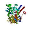
| ||||||||
|---|---|---|---|---|---|---|---|---|---|
| 1 | 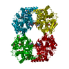
| ||||||||
| Unit cell |
|
-Components
-Protein , 1 types, 1 molecules A
| #1: Protein | Mass: 55294.203 Da / Num. of mol.: 1 Source method: isolated from a genetically manipulated source Source: (gene. exp.) Homo sapiens (human) / Gene: NT5C2, NT5B, NT5CP, PNT5 / Plasmid: pET28 / Production host:  |
|---|
-Non-polymers , 5 types, 310 molecules 


| #2: Chemical | ChemComp-GOL /  Mass: 92.094 Da / Num. of mol.: 9 / Source method: obtained synthetically / Formula: C3H8O3 Mass: 92.094 Da / Num. of mol.: 9 / Source method: obtained synthetically / Formula: C3H8O3#3: Chemical | ChemComp-PO4 / |  Mass: 94.971 Da / Num. of mol.: 1 / Source method: obtained synthetically / Formula: PO4 Mass: 94.971 Da / Num. of mol.: 1 / Source method: obtained synthetically / Formula: PO4#4: Chemical |  Mass: 24.305 Da / Num. of mol.: 2 / Source method: obtained synthetically / Formula: Mg Mass: 24.305 Da / Num. of mol.: 2 / Source method: obtained synthetically / Formula: Mg#5: Chemical | ChemComp-DTP / |  Mass: 491.182 Da / Num. of mol.: 1 / Source method: obtained synthetically / Formula: C10H16N5O12P3 Mass: 491.182 Da / Num. of mol.: 1 / Source method: obtained synthetically / Formula: C10H16N5O12P3#6: Water | ChemComp-HOH / | Mass: 18.015 Da / Num. of mol.: 297 / Source method: isolated from a natural source / Formula: H2O |
|---|
-Experimental details
-Experiment
| Experiment | Method: X-RAY DIFFRACTION / Number of used crystals: 1 |
|---|
- Sample preparation
| Crystal | Density Matthews: 3.43 Å3/Da / Density % sol: 64.13 % |
|---|---|
| Crystal grow | Temperature: 293 K / Method: vapor diffusion Details: 0.1 M MES/imidazole pH 6.5; 0.03 M of each divalent cation; 10% w/v PEG 8000, 20% v/v ethylene glycol (Morpheus condition A2) |
-Data collection
| Diffraction | Mean temperature: 100 K |
|---|---|
| Diffraction source | Source: SYNCHROTRON / Site: BESSY  / Beamline: 14.1 / Wavelength: 0.918409 Å / Beamline: 14.1 / Wavelength: 0.918409 Å |
| Detector | Type: DECTRIS PILATUS 6M / Detector: PIXEL / Date: Apr 30, 2015 |
| Radiation | Protocol: SINGLE WAVELENGTH / Monochromatic (M) / Laue (L): M / Scattering type: x-ray |
| Radiation wavelength | Wavelength: 0.918409 Å / Relative weight: 1 |
| Reflection | Resolution: 1.736→48.99 Å / Num. obs: 77607 / % possible obs: 98.8 % / Redundancy: 6.5 % / Biso Wilson estimate: 29.7 Å2 / CC1/2: 0.998 / Rmerge(I) obs: 0.093 / Rrim(I) all: 0.101 / Net I/av σ(I): 13.55 / Net I/σ(I): 13.6 |
| Reflection shell | Resolution: 1.74→1.84 Å / Redundancy: 6.21 % / Rmerge(I) obs: 0.83 / Mean I/σ(I) obs: 1.88 / Num. unique obs: 11899 / CC1/2: 0.823 / Rrim(I) all: 0.905 / % possible all: 94.6 |
- Processing
| Software |
| ||||||||||||||||||||||||||||||||||||||||||||||||||||||||||||||||||||||||||||||||||||||||||||||||||||||||||||||||||||||||||||||||||||||||||||||||||||||||||||||||||||||||||||||||||||||||||||||||||||||||||||||||||||||||||||||||||||||||||||||||||||||||||||||||||||||||||||||||||||||||||||||||||||||||||||||||||||||||||||||||||||||||||||||||||||||||||||||||||||||||||||||||||||||||||||||||||||||||||||||||
|---|---|---|---|---|---|---|---|---|---|---|---|---|---|---|---|---|---|---|---|---|---|---|---|---|---|---|---|---|---|---|---|---|---|---|---|---|---|---|---|---|---|---|---|---|---|---|---|---|---|---|---|---|---|---|---|---|---|---|---|---|---|---|---|---|---|---|---|---|---|---|---|---|---|---|---|---|---|---|---|---|---|---|---|---|---|---|---|---|---|---|---|---|---|---|---|---|---|---|---|---|---|---|---|---|---|---|---|---|---|---|---|---|---|---|---|---|---|---|---|---|---|---|---|---|---|---|---|---|---|---|---|---|---|---|---|---|---|---|---|---|---|---|---|---|---|---|---|---|---|---|---|---|---|---|---|---|---|---|---|---|---|---|---|---|---|---|---|---|---|---|---|---|---|---|---|---|---|---|---|---|---|---|---|---|---|---|---|---|---|---|---|---|---|---|---|---|---|---|---|---|---|---|---|---|---|---|---|---|---|---|---|---|---|---|---|---|---|---|---|---|---|---|---|---|---|---|---|---|---|---|---|---|---|---|---|---|---|---|---|---|---|---|---|---|---|---|---|---|---|---|---|---|---|---|---|---|---|---|---|---|---|---|---|---|---|---|---|---|---|---|---|---|---|---|---|---|---|---|---|---|---|---|---|---|---|---|---|---|---|---|---|---|---|---|---|---|---|---|---|---|---|---|---|---|---|---|---|---|---|---|---|---|---|---|---|---|---|---|---|---|---|---|---|---|---|---|---|---|---|---|---|---|---|---|---|---|---|---|---|---|---|---|---|---|---|---|---|---|---|---|---|---|---|---|---|---|---|---|---|---|---|---|---|---|---|---|---|---|---|---|---|---|---|---|---|---|---|---|---|---|---|---|---|---|---|---|---|---|---|---|---|---|---|---|---|---|---|---|---|---|---|
| Refinement | Method to determine structure: MOLECULAR REPLACEMENT Starting model: 5K7Y Resolution: 1.74→48.99 Å / Cor.coef. Fo:Fc: 0.962 / Cor.coef. Fo:Fc free: 0.951 / Cross valid method: THROUGHOUT / σ(F): 0 / ESU R: 0.088 / ESU R Free: 0.088 Details: HYDROGENS HAVE BEEN ADDED IN THE RIDING POSITIONS U VALUES : WITH TLS ADDED
| ||||||||||||||||||||||||||||||||||||||||||||||||||||||||||||||||||||||||||||||||||||||||||||||||||||||||||||||||||||||||||||||||||||||||||||||||||||||||||||||||||||||||||||||||||||||||||||||||||||||||||||||||||||||||||||||||||||||||||||||||||||||||||||||||||||||||||||||||||||||||||||||||||||||||||||||||||||||||||||||||||||||||||||||||||||||||||||||||||||||||||||||||||||||||||||||||||||||||||||||||
| Solvent computation | Ion probe radii: 0.8 Å / Shrinkage radii: 0.8 Å / VDW probe radii: 1.2 Å | ||||||||||||||||||||||||||||||||||||||||||||||||||||||||||||||||||||||||||||||||||||||||||||||||||||||||||||||||||||||||||||||||||||||||||||||||||||||||||||||||||||||||||||||||||||||||||||||||||||||||||||||||||||||||||||||||||||||||||||||||||||||||||||||||||||||||||||||||||||||||||||||||||||||||||||||||||||||||||||||||||||||||||||||||||||||||||||||||||||||||||||||||||||||||||||||||||||||||||||||||
| Displacement parameters | Biso max: 105.74 Å2 / Biso mean: 29.152 Å2 / Biso min: 3.04 Å2
| ||||||||||||||||||||||||||||||||||||||||||||||||||||||||||||||||||||||||||||||||||||||||||||||||||||||||||||||||||||||||||||||||||||||||||||||||||||||||||||||||||||||||||||||||||||||||||||||||||||||||||||||||||||||||||||||||||||||||||||||||||||||||||||||||||||||||||||||||||||||||||||||||||||||||||||||||||||||||||||||||||||||||||||||||||||||||||||||||||||||||||||||||||||||||||||||||||||||||||||||||
| Refinement step | Cycle: final / Resolution: 1.74→48.99 Å
| ||||||||||||||||||||||||||||||||||||||||||||||||||||||||||||||||||||||||||||||||||||||||||||||||||||||||||||||||||||||||||||||||||||||||||||||||||||||||||||||||||||||||||||||||||||||||||||||||||||||||||||||||||||||||||||||||||||||||||||||||||||||||||||||||||||||||||||||||||||||||||||||||||||||||||||||||||||||||||||||||||||||||||||||||||||||||||||||||||||||||||||||||||||||||||||||||||||||||||||||||
| Refine LS restraints |
| ||||||||||||||||||||||||||||||||||||||||||||||||||||||||||||||||||||||||||||||||||||||||||||||||||||||||||||||||||||||||||||||||||||||||||||||||||||||||||||||||||||||||||||||||||||||||||||||||||||||||||||||||||||||||||||||||||||||||||||||||||||||||||||||||||||||||||||||||||||||||||||||||||||||||||||||||||||||||||||||||||||||||||||||||||||||||||||||||||||||||||||||||||||||||||||||||||||||||||||||||
| LS refinement shell | Resolution: 1.736→1.781 Å / Rfactor Rfree error: 0 / Total num. of bins used: 20
| ||||||||||||||||||||||||||||||||||||||||||||||||||||||||||||||||||||||||||||||||||||||||||||||||||||||||||||||||||||||||||||||||||||||||||||||||||||||||||||||||||||||||||||||||||||||||||||||||||||||||||||||||||||||||||||||||||||||||||||||||||||||||||||||||||||||||||||||||||||||||||||||||||||||||||||||||||||||||||||||||||||||||||||||||||||||||||||||||||||||||||||||||||||||||||||||||||||||||||||||||
| Refinement TLS params. | Method: refined / Refine-ID: X-RAY DIFFRACTION
| ||||||||||||||||||||||||||||||||||||||||||||||||||||||||||||||||||||||||||||||||||||||||||||||||||||||||||||||||||||||||||||||||||||||||||||||||||||||||||||||||||||||||||||||||||||||||||||||||||||||||||||||||||||||||||||||||||||||||||||||||||||||||||||||||||||||||||||||||||||||||||||||||||||||||||||||||||||||||||||||||||||||||||||||||||||||||||||||||||||||||||||||||||||||||||||||||||||||||||||||||
| Refinement TLS group |
|
