 Movie
Movie Controller

[English] 日本語
 Yorodumi
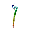
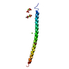
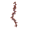
- PDB-5omd: Crystal structure of S. cerevisiae Ddc2 N-terminal coiled-coil domain -
+ Open data

- Basic information
| Entry | Database: PDB / ID: 5omd | ||||||
|---|---|---|---|---|---|---|---|
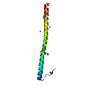
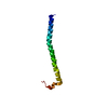
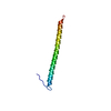
| Title | Crystal structure of S. cerevisiae Ddc2 N-terminal coiled-coil domain | ||||||
 Components Components | DNA damage checkpoint protein LCD1 | ||||||
 Keywords Keywords | PROTEIN BINDING / coiled-coil / dimerization | ||||||
| Function / homology |  Function and homology information Function and homology informationATR-ATRIP complex / regulation of double-strand break repair / nucleobase-containing compound metabolic process / nuclear chromosome / telomere maintenance via telomerase / DNA damage checkpoint signaling / establishment of protein localization / chromatin organization / damaged DNA binding / DNA repair ...ATR-ATRIP complex / regulation of double-strand break repair / nucleobase-containing compound metabolic process / nuclear chromosome / telomere maintenance via telomerase / DNA damage checkpoint signaling / establishment of protein localization / chromatin organization / damaged DNA binding / DNA repair / nucleus / cytoplasm Similarity search - Function | ||||||
| Biological species |  | ||||||
| Method |  X-RAY DIFFRACTION / SYNCHROTRON / MOLECULAR REPLACEMENT / Resolution: 2.1 Å X-RAY DIFFRACTION / SYNCHROTRON / MOLECULAR REPLACEMENT / Resolution: 2.1 Å | ||||||
 Authors Authors | Deshpande, I. / Seeber, A. / Shimada, K. / Keusch, J.J. / Gut, H. / Gasser, S.M. | ||||||
 Citation Citation | Journal: Mol. Cell / Year: 2017 Title: Structural Basis of Mec1-Ddc2-RPA Assembly and Activation on Single-Stranded DNA at Sites of Damage. Authors: Deshpande, I. / Seeber, A. / Shimada, K. / Keusch, J.J. / Gut, H. / Gasser, S.M. | ||||||
| History |
|
- Structure visualization
| Structure viewer | Molecule: MolmilJmol/JSmol |
|---|
- Downloads & links
-Download
| PDBx/mmCIF format | 5omd.cif.gz | 39.8 KB | Display | PDBx/mmCIF format |
|---|---|---|---|---|
| PDB format | pdb5omd.ent.gz | 27.3 KB | Display | PDB format |
| PDBx/mmJSON format | 5omd.json.gz | Tree view | PDBx/mmJSON format | |
| Others |  Other downloads Other downloads |
-Validation report
| Arichive directory | https://data.pdbj.org/pub/pdb/validation_reports/om/5omdftp://data.pdbj.org/pub/pdb/validation_reports/om/5omd | HTTPS FTP |
|---|
-Related structure data
| Related structure data |  5ombC  5omcC 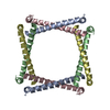 1a92S S: Starting model for refinement C: citing same article ( |
|---|---|
| Similar structure data |

-Links
 PDBj
PDBj- Assembly
| Deposited unit | 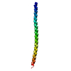
| ||||||||||||
|---|---|---|---|---|---|---|---|---|---|---|---|---|---|
| 1 | 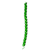
| ||||||||||||
| Unit cell |
| ||||||||||||
| Components on special symmetry positions |
|
-Components
| #1: Protein | Mass: 7733.979 Da / Num. of mol.: 1 / Fragment: UNP residues 73-136 Source method: isolated from a genetically manipulated source Source: (gene. exp.) Gene: LCD1, DDC2, PIE1, YDR499W / Plasmid: pOPINF / Production host:  |
|---|---|
| #2: Water | ChemComp-HOH /  Mass: 18.015 Da / Num. of mol.: 45 / Source method: isolated from a natural source / Formula: H2O Mass: 18.015 Da / Num. of mol.: 45 / Source method: isolated from a natural source / Formula: H2O |
-Experimental details
-Experiment
| Experiment | Method: X-RAY DIFFRACTION / Number of used crystals: 1 |
|---|
- Sample preparation
| Crystal | Density Matthews: 2.32 Å3/Da / Density % sol: 46.98 % |
|---|---|
| Crystal grow | Temperature: 293 K / Method: vapor diffusion, sitting drop / pH: 8.5 / Details: 0.1 M Tris-HCl, 24% PEG 400 |
-Data collection
| Diffraction | Mean temperature: 100 K |
|---|---|
| Diffraction source | Source: SYNCHROTRON / Site: SLS  / Beamline: X06DA / Wavelength: 1.00005 Å / Beamline: X06DA / Wavelength: 1.00005 Å |
| Detector | Type: DECTRIS PILATUS 2M / Detector: PIXEL / Date: May 6, 2013 |
| Radiation | Protocol: SINGLE WAVELENGTH / Monochromatic (M) / Laue (L): M / Scattering type: x-ray |
| Radiation wavelength | Wavelength: 1.00005 Å / Relative weight: 1 |
| Reflection | Resolution: 2.1→50 Å / Num. obs: 4644 / % possible obs: 99.8 % / Redundancy: 6.08 % / Biso Wilson estimate: 35.69 Å2 / CC1/2: 0.999 / Rsym value: 0.09 / Net I/σ(I): 13.23 |
| Reflection shell | Resolution: 2.1→2.15 Å / Redundancy: 5.72 % / Mean I/σ(I) obs: 2.19 / Num. unique obs: 330 / CC1/2: 0.706 / Rsym value: 0.856 / % possible all: 98.5 |
- Processing
| Software |
| ||||||||||||||||||||||||||||||||||||||||||||||||||||||||||||||||||||||||||||||||||||||||||||||||||||||||||||||||||
|---|---|---|---|---|---|---|---|---|---|---|---|---|---|---|---|---|---|---|---|---|---|---|---|---|---|---|---|---|---|---|---|---|---|---|---|---|---|---|---|---|---|---|---|---|---|---|---|---|---|---|---|---|---|---|---|---|---|---|---|---|---|---|---|---|---|---|---|---|---|---|---|---|---|---|---|---|---|---|---|---|---|---|---|---|---|---|---|---|---|---|---|---|---|---|---|---|---|---|---|---|---|---|---|---|---|---|---|---|---|---|---|---|---|---|---|
| Refinement | Method to determine structure: MOLECULAR REPLACEMENT Starting model: 1A92 Resolution: 2.1→38.98 Å / Cor.coef. Fo:Fc: 0.9406 / Cor.coef. Fo:Fc free: 0.9456 / SU R Cruickshank DPI: 0.231 / Cross valid method: THROUGHOUT / σ(F): 0 / SU R Blow DPI: 0.269 / SU Rfree Blow DPI: 0.209 / SU Rfree Cruickshank DPI: 0.196
| ||||||||||||||||||||||||||||||||||||||||||||||||||||||||||||||||||||||||||||||||||||||||||||||||||||||||||||||||||
| Displacement parameters | Biso mean: 51.81 Å2
| ||||||||||||||||||||||||||||||||||||||||||||||||||||||||||||||||||||||||||||||||||||||||||||||||||||||||||||||||||
| Refine analyze | Luzzati coordinate error obs: 0.347 Å | ||||||||||||||||||||||||||||||||||||||||||||||||||||||||||||||||||||||||||||||||||||||||||||||||||||||||||||||||||
| Refinement step | Cycle: LAST / Resolution: 2.1→38.98 Å
| ||||||||||||||||||||||||||||||||||||||||||||||||||||||||||||||||||||||||||||||||||||||||||||||||||||||||||||||||||
| Refine LS restraints |
| ||||||||||||||||||||||||||||||||||||||||||||||||||||||||||||||||||||||||||||||||||||||||||||||||||||||||||||||||||
| LS refinement shell | Resolution: 2.1→2.35 Å / Total num. of bins used: 5
| ||||||||||||||||||||||||||||||||||||||||||||||||||||||||||||||||||||||||||||||||||||||||||||||||||||||||||||||||||
| Refinement TLS params. | Method: refined / Origin x: 28.4228 Å / Origin y: 5.3861 Å / Origin z: 14.1176 Å
| ||||||||||||||||||||||||||||||||||||||||||||||||||||||||||||||||||||||||||||||||||||||||||||||||||||||||||||||||||
| Refinement TLS group | Selection details: { A|* } |
