 ムービー
ムービー コントローラー


+ データを開く

- 基本情報
基本情報
| 登録情報 | データベース: PDB / ID: 5ofo | ||||||
|---|---|---|---|---|---|---|---|
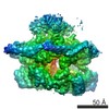
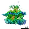
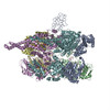
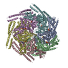
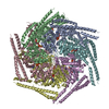
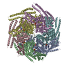
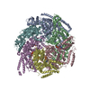
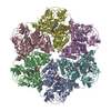
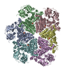
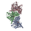
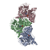
| タイトル | Cryo EM structure of the E. coli disaggregase ClpB (BAP form, DWB mutant), in the ATPgammaS state, bound to the model substrate casein | ||||||
 要素 要素 | Chaperone protein ClpB,ATP-dependent Clp protease ATP-binding subunit ClpA,Chaperone protein ClpB | ||||||
 キーワード キーワード | CHAPERONE / disaggregase / ClpB / AAA | ||||||
| 機能・相同性 |  機能・相同性情報 機能・相同性情報endopeptidase Clp complex / ATP-dependent peptidase activity / protein quality control for misfolded or incompletely synthesized proteins / protein unfolding / cellular response to heat / response to heat / protein refolding / response to oxidative stress / ATP hydrolysis activity / proteolysis ...endopeptidase Clp complex / ATP-dependent peptidase activity / protein quality control for misfolded or incompletely synthesized proteins / protein unfolding / cellular response to heat / response to heat / protein refolding / response to oxidative stress / ATP hydrolysis activity / proteolysis / ATP binding / identical protein binding / membrane / cytosol / cytoplasm 類似検索 - 分子機能 | ||||||
| 生物種 |  | ||||||
| 手法 | 電子顕微鏡法 / 単粒子再構成法 / クライオ電子顕微鏡法 / 解像度: 4.6 Å | ||||||
 データ登録者 データ登録者 | Deville, C. / Carroni, M. / Franke, K.B. / Topf, M. / Bukau, B. / Mogk, A. / Saibil, H.R. | ||||||
 引用 引用 | ジャーナル: Sci Adv / 年: 2017 タイトル: Structural pathway of regulated substrate transfer and threading through an Hsp100 disaggregase. 著者: Célia Deville / Marta Carroni / Kamila B Franke / Maya Topf / Bernd Bukau / Axel Mogk / Helen R Saibil /   要旨: Refolding aggregated proteins is essential in combating cellular proteotoxic stress. Together with Hsp70, Hsp100 chaperones, including ClpB, form a powerful disaggregation machine that threads ...Refolding aggregated proteins is essential in combating cellular proteotoxic stress. Together with Hsp70, Hsp100 chaperones, including ClpB, form a powerful disaggregation machine that threads aggregated polypeptides through the central pore of tandem adenosine triphosphatase (ATPase) rings. To visualize protein disaggregation, we determined cryo-electron microscopy structures of inactive and substrate-bound ClpB in the presence of adenosine 5'--(3-thiotriphosphate), revealing closed AAA+ rings with a pronounced seam. In the substrate-free state, a marked gradient of resolution, likely corresponding to mobility, spans across the AAA+ rings with a dynamic hotspot at the seam. On the seam side, the coiled-coil regulatory domains are locked in a horizontal, inactive orientation. On the opposite side, the regulatory domains are accessible for Hsp70 binding, substrate targeting, and activation. In the presence of the model substrate casein, the polypeptide threads through the entire pore channel and increased nucleotide occupancy correlates with higher ATPase activity. Substrate-induced domain displacements indicate a pathway of regulated substrate transfer from Hsp70 to the ClpB pore, inside which a spiral of loops contacts the substrate. The seam pore loops undergo marked displacements, along with ordering of the regulatory domains. These asymmetric movements suggest a mechanism for ATPase activation and substrate threading during disaggregation. | ||||||
| 履歴 |
|
- 構造の表示
構造の表示
| ムービー |
ムービービューア |
|---|---|
| 構造ビューア | 分子: MolmilJmol/JSmol |
- ダウンロードとリンク
-ダウンロード
| PDBx/mmCIF形式 | 5ofo.cif.gz | 718.1 KB | 表示 | PDBx/mmCIF形式 |
|---|---|---|---|---|
| PDB形式 | pdb5ofo.ent.gz | 561.3 KB | 表示 | PDB形式 |
| PDBx/mmJSON形式 | 5ofo.json.gz | ツリー表示 | PDBx/mmJSON形式 | |
| その他 |  その他のダウンロード その他のダウンロード |
-検証レポート
| 文書・要旨 | 5ofo_validation.pdf.gz | 2 MB | 表示 | wwPDB検証レポート |
|---|---|---|---|---|
| 文書・詳細版 | 5ofo_full_validation.pdf.gz | 2.1 MB | 表示 | |
| XML形式データ | 5ofo_validation.xml.gz | 125.6 KB | 表示 | |
| CIF形式データ | 5ofo_validation.cif.gz | 186.2 KB | 表示 | |
| アーカイブディレクトリ | https://data.pdbj.org/pub/pdb/validation_reports/of/5ofoftp://data.pdbj.org/pub/pdb/validation_reports/of/5ofo | HTTPS FTP |
-関連構造データ
-リンク
 PDBj
PDBj


- 集合体
集合体
| 登録構造単位 | 
|
|---|---|
| 1 |
|
-要素
| #1: タンパク質 | 分子量: 97288.914 Da / 分子数: 6 / 由来タイプ: 組換発現 由来: (組換発現) 株: K12 遺伝子: clpB, htpM, b2592, JW2573, clpA, lopD, b0882, JW0866 発現宿主: #2: 化合物 | ChemComp-AGS /   分子量: 523.247 Da / 分子数: 11 / 由来タイプ: 合成 / 式: C10H16N5O12P3S 分子量: 523.247 Da / 分子数: 11 / 由来タイプ: 合成 / 式: C10H16N5O12P3Sコメント: ATP-gamma-S, エネルギー貯蔵分子類似体*YM |
|---|
-実験情報
-実験
| 実験 | 手法: 電子顕微鏡法 |
|---|---|
| EM実験 | 試料の集合状態: PARTICLE / 3次元再構成法: 単粒子再構成法 |
- 試料調製
試料調製
| 構成要素 | 名称: ClpB, BAP form, double walker B mutant / タイプ: COMPLEX / Entity ID: #1 / 由来: RECOMBINANT | ||||||||||||||||||||||||||||||
|---|---|---|---|---|---|---|---|---|---|---|---|---|---|---|---|---|---|---|---|---|---|---|---|---|---|---|---|---|---|---|---|
| 分子量 | 値: 0.57 MDa / 実験値: NO | ||||||||||||||||||||||||||||||
| 由来(天然) | 生物種: | ||||||||||||||||||||||||||||||
| 由来(組換発現) | 生物種: | ||||||||||||||||||||||||||||||
| 緩衝液 | pH: 7.4 | ||||||||||||||||||||||||||||||
| 緩衝液成分 |
| ||||||||||||||||||||||||||||||
| 試料 | 濃度: 0.7 mg/ml / 包埋: NO / シャドウイング: NO / 染色: NO / 凍結: YES | ||||||||||||||||||||||||||||||
| 試料支持 | グリッドの材料: COPPER / グリッドのサイズ: 400 divisions/in. / グリッドのタイプ: Agar lacey carbon | ||||||||||||||||||||||||||||||
| 急速凍結 | 装置: FEI VITROBOT MARK III / 凍結剤: ETHANE / 凍結前の試料温度: 295 K |
- 電子顕微鏡撮影
| 実験機器 |  モデル: Titan Krios / 画像提供: FEI Company |
|---|---|
| 顕微鏡 | モデル: FEI TITAN KRIOS |
| 電子銃 | 電子線源:  FIELD EMISSION GUN / 加速電圧: 300 kV / 照射モード: FLOOD BEAM FIELD EMISSION GUN / 加速電圧: 300 kV / 照射モード: FLOOD BEAM |
| 電子レンズ | モード: BRIGHT FIELD / 最大 デフォーカス(公称値): 3000 nm / 最小 デフォーカス(公称値): 1000 nm |
| 試料ホルダ | 凍結剤: NITROGEN 試料ホルダーモデル: FEI TITAN KRIOS AUTOGRID HOLDER |
| 撮影 | 電子線照射量: 1 e/Å2 / 検出モード: COUNTING フィルム・検出器のモデル: GATAN K2 SUMMIT (4k x 4k) |
| 画像スキャン | サンプリングサイズ: 5 µm / 動画フレーム数/画像: 50 |
- 解析
解析
| ソフトウェア | 名称: PHENIX / バージョン: 1.11.1_2575: / 分類: 精密化 | |||||||||||||||||||||||||||||||||||||||||||||||||||||||
|---|---|---|---|---|---|---|---|---|---|---|---|---|---|---|---|---|---|---|---|---|---|---|---|---|---|---|---|---|---|---|---|---|---|---|---|---|---|---|---|---|---|---|---|---|---|---|---|---|---|---|---|---|---|---|---|---|
| EMソフトウェア |
| |||||||||||||||||||||||||||||||||||||||||||||||||||||||
| CTF補正 | タイプ: PHASE FLIPPING AND AMPLITUDE CORRECTION | |||||||||||||||||||||||||||||||||||||||||||||||||||||||
| 粒子像の選択 | 選択した粒子像数: 500000 | |||||||||||||||||||||||||||||||||||||||||||||||||||||||
| 対称性 | 点対称性: C1 (非対称) | |||||||||||||||||||||||||||||||||||||||||||||||||||||||
| 3次元再構成 | 解像度: 4.6 Å / 解像度の算出法: FSC 0.143 CUT-OFF / 粒子像の数: 230000 / 対称性のタイプ: POINT | |||||||||||||||||||||||||||||||||||||||||||||||||||||||
| 原子モデル構築 | プロトコル: FLEXIBLE FIT / 空間: REAL / Target criteria: Local correlation coefficient 詳細: Initial models of the monomer were generated using MODELLER v9.17 with previously determined crystal structures of ClpB or ClpB domains as templates (PDB ids 4CIU, 1QVR, 4HSE and 4LJ9). The ...詳細: Initial models of the monomer were generated using MODELLER v9.17 with previously determined crystal structures of ClpB or ClpB domains as templates (PDB ids 4CIU, 1QVR, 4HSE and 4LJ9). The crystal structure of E. coli ClpB (4CIU) was used as a main template, the crystal structure of T. thermophilus (1QVR) was used to model positions of the NTD and the crystal structures of AAA1 and AAA2 of E. coli ClpB (4HSE and 4LJ9) were used to model the pore loops disordered in other crystal structures. Initial rigid body fitting of the monomers in the map was manually done in Chimera using the Fit-in-Map tool. iMODFIT was used for fitting involving large domain motions. FlexEM was then used for refinement of secondary structures and loops in the map. Quality and improvement of the fit was assessed with TEMPy using the Segment based Manders Overlap Coefficient (SMOC) scores and segment based cross correlation scores. Ridig body fitting of ATPgammaS (structure extracted from PDB id 3EIH) into the nucleotide biding sides was manually done in Chimera using the Fit-in-Map tool. The target map for fitting of ATPgammaS was the difference map between experimental map and map generated using the nucleotide-free protein model revealing nucleotide densities. A round of real-space refinement was performed in phenix using energy minimization to fix the model's geometry and clashes. | |||||||||||||||||||||||||||||||||||||||||||||||||||||||
| 拘束条件 |
|
