 Movie
Movie Controller

[English] 日本語
 Yorodumi
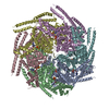
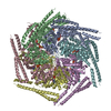
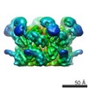
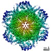
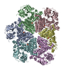
- PDB-5og1: Cryo EM structure of the E. coli disaggregase ClpB (BAP form, DWB... -
+ Open data

- Basic information
| Entry | Database: PDB / ID: 5og1 | ||||||||||||||||||||||||
|---|---|---|---|---|---|---|---|---|---|---|---|---|---|---|---|---|---|---|---|---|---|---|---|---|---|
| Title | Cryo EM structure of the E. coli disaggregase ClpB (BAP form, DWB mutant), in the ATPgammaS state | ||||||||||||||||||||||||
 Components Components | Chaperone protein ClpB,ATP-dependent Clp protease ATP-binding subunit ClpA,Chaperone protein ClpB | ||||||||||||||||||||||||
 Keywords Keywords | CHAPERONE / disaggregase / ClpB / AAA | ||||||||||||||||||||||||
| Function / homology |  Function and homology information Function and homology informationendopeptidase Clp complex / ATP-dependent peptidase activity / protein quality control for misfolded or incompletely synthesized proteins / protein unfolding / cellular response to heat / response to heat / protein refolding / response to oxidative stress / ATP hydrolysis activity / proteolysis ...endopeptidase Clp complex / ATP-dependent peptidase activity / protein quality control for misfolded or incompletely synthesized proteins / protein unfolding / cellular response to heat / response to heat / protein refolding / response to oxidative stress / ATP hydrolysis activity / proteolysis / ATP binding / membrane / identical protein binding / cytoplasm / cytosol Similarity search - Function | ||||||||||||||||||||||||
| Biological species |  | ||||||||||||||||||||||||
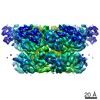
| Method | ELECTRON MICROSCOPY / single particle reconstruction / cryo EM / Resolution: 4.5 Å | ||||||||||||||||||||||||
 Authors Authors | Deville, C. / Carroni, M. / Franke, K.B. / Topf, M. / Bukau, B. / Mogk, A. / Saibil, H.R. | ||||||||||||||||||||||||
| Funding support |  United Kingdom, United Kingdom,  Germany, 7items Germany, 7items
| ||||||||||||||||||||||||
 Citation Citation | Journal: Sci Adv / Year: 2017 Title: Structural pathway of regulated substrate transfer and threading through an Hsp100 disaggregase. Authors: Célia Deville / Marta Carroni / Kamila B Franke / Maya Topf / Bernd Bukau / Axel Mogk / Helen R Saibil / Abstract: Refolding aggregated proteins is essential in combating cellular proteotoxic stress. Together with Hsp70, Hsp100 chaperones, including ClpB, form a powerful disaggregation machine that threads ...Refolding aggregated proteins is essential in combating cellular proteotoxic stress. Together with Hsp70, Hsp100 chaperones, including ClpB, form a powerful disaggregation machine that threads aggregated polypeptides through the central pore of tandem adenosine triphosphatase (ATPase) rings. To visualize protein disaggregation, we determined cryo-electron microscopy structures of inactive and substrate-bound ClpB in the presence of adenosine 5'--(3-thiotriphosphate), revealing closed AAA+ rings with a pronounced seam. In the substrate-free state, a marked gradient of resolution, likely corresponding to mobility, spans across the AAA+ rings with a dynamic hotspot at the seam. On the seam side, the coiled-coil regulatory domains are locked in a horizontal, inactive orientation. On the opposite side, the regulatory domains are accessible for Hsp70 binding, substrate targeting, and activation. In the presence of the model substrate casein, the polypeptide threads through the entire pore channel and increased nucleotide occupancy correlates with higher ATPase activity. Substrate-induced domain displacements indicate a pathway of regulated substrate transfer from Hsp70 to the ClpB pore, inside which a spiral of loops contacts the substrate. The seam pore loops undergo marked displacements, along with ordering of the regulatory domains. These asymmetric movements suggest a mechanism for ATPase activation and substrate threading during disaggregation. | ||||||||||||||||||||||||
| History |
|
- Structure visualization
| Movie |
Movie viewer |
|---|---|
| Structure viewer | Molecule: MolmilJmol/JSmol |
- Downloads & links
-Download
| PDBx/mmCIF format | 5og1.cif.gz | 691.6 KB | Display | PDBx/mmCIF format |
|---|---|---|---|---|
| PDB format | pdb5og1.ent.gz | 535.1 KB | Display | PDB format |
| PDBx/mmJSON format | 5og1.json.gz | Tree view | PDBx/mmJSON format | |
| Others |  Other downloads Other downloads |
-Validation report
| Arichive directory | https://data.pdbj.org/pub/pdb/validation_reports/og/5og1ftp://data.pdbj.org/pub/pdb/validation_reports/og/5og1 | HTTPS FTP |
|---|
-Related structure data
| Related structure data | 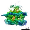 3777MC 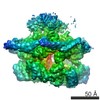 3776C 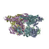 5ofoC M: map data used to model this data C: citing same article ( |
|---|---|
| Similar structure data |

-Links
 PDBj
PDBj


- Assembly
| Deposited unit | 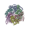
|
|---|---|
| 1 |
|
-Components
| #1: Protein | Mass: 97288.914 Da / Num. of mol.: 6 Source method: isolated from a genetically manipulated source Source: (gene. exp.) Strain: K12 / Gene: clpB, htpM, b2592, JW2573, clpA, lopD, b0882, JW0866 / Production host: #2: Chemical | ChemComp-AGS /   Mass: 523.247 Da / Num. of mol.: 7 / Source method: obtained synthetically / Formula: C10H16N5O12P3S / Comment: ATP-gamma-S, energy-carrying molecule analogue*YM Mass: 523.247 Da / Num. of mol.: 7 / Source method: obtained synthetically / Formula: C10H16N5O12P3S / Comment: ATP-gamma-S, energy-carrying molecule analogue*YM |
|---|
-Experimental details
-Experiment
| Experiment | Method: ELECTRON MICROSCOPY |
|---|---|
| EM experiment | Aggregation state: PARTICLE / 3D reconstruction method: single particle reconstruction |
- Sample preparation
| Component | Name: ClpB (BAP form, double walker B mutant) homohexamer in the ATPgammaS state Type: COMPLEX / Entity ID: #1-#4 / Source: RECOMBINANT | ||||||||||||||||||||||||||||||
|---|---|---|---|---|---|---|---|---|---|---|---|---|---|---|---|---|---|---|---|---|---|---|---|---|---|---|---|---|---|---|---|
| Molecular weight | Value: 0.57 MDa / Experimental value: NO | ||||||||||||||||||||||||||||||
| Source (natural) | Organism: | ||||||||||||||||||||||||||||||
| Source (recombinant) | Organism: | ||||||||||||||||||||||||||||||
| Buffer solution | pH: 7.4 | ||||||||||||||||||||||||||||||
| Buffer component |
| ||||||||||||||||||||||||||||||
| Specimen | Conc.: 0.7 mg/ml / Embedding applied: NO / Shadowing applied: NO / Staining applied: NO / Vitrification applied: YES | ||||||||||||||||||||||||||||||
| Specimen support | Grid material: COPPER / Grid mesh size: 400 divisions/in. / Grid type: Agar lacey carbon | ||||||||||||||||||||||||||||||
| Vitrification | Instrument: FEI VITROBOT MARK III / Cryogen name: ETHANE / Humidity: 90 % / Chamber temperature: 295 K |
- Electron microscopy imaging
| Experimental equipment |  Model: Titan Krios / Image courtesy: FEI Company |
|---|---|
| Microscopy | Model: FEI TITAN KRIOS |
| Electron gun | Electron source:  FIELD EMISSION GUN / Accelerating voltage: 300 kV / Illumination mode: FLOOD BEAM FIELD EMISSION GUN / Accelerating voltage: 300 kV / Illumination mode: FLOOD BEAM |
| Electron lens | Mode: BRIGHT FIELD / Nominal defocus max: 3000 nm / Nominal defocus min: 1000 nm |
| Specimen holder | Cryogen: NITROGEN / Specimen holder model: FEI TITAN KRIOS AUTOGRID HOLDER |
| Image recording | Electron dose: 1 e/Å2 / Detector mode: COUNTING / Film or detector model: GATAN K2 SUMMIT (4k x 4k) |
| Image scans | Sampling size: 5 µm / Movie frames/image: 50 |
- Processing
| EM software |
| ||||||||||||||||||||||||||||||||||||||||||||
|---|---|---|---|---|---|---|---|---|---|---|---|---|---|---|---|---|---|---|---|---|---|---|---|---|---|---|---|---|---|---|---|---|---|---|---|---|---|---|---|---|---|---|---|---|---|
| CTF correction | Type: PHASE FLIPPING AND AMPLITUDE CORRECTION | ||||||||||||||||||||||||||||||||||||||||||||
| Particle selection | Num. of particles selected: 250000 | ||||||||||||||||||||||||||||||||||||||||||||
| Symmetry | Point symmetry: C1 (asymmetric) | ||||||||||||||||||||||||||||||||||||||||||||
| 3D reconstruction | Resolution: 4.5 Å / Resolution method: FSC 0.143 CUT-OFF / Num. of particles: 60000 / Symmetry type: POINT | ||||||||||||||||||||||||||||||||||||||||||||
| Atomic model building | Protocol: FLEXIBLE FIT / Space: REAL / Target criteria: local correlation coeficient Details: Initial models of the BAPDWB monomer were generated using MODELLER v9.17 with previously determined crystal structures of ClpB or ClpB domains as templates (PDB ids 4CIU, 1QVR, 4HSE and 4LJ9) ...Details: Initial models of the BAPDWB monomer were generated using MODELLER v9.17 with previously determined crystal structures of ClpB or ClpB domains as templates (PDB ids 4CIU, 1QVR, 4HSE and 4LJ9). The crystal structure of E. coli ClpB (4CIU) was used as a main template, the crystal structure of T. thermophilus (1QVR) was used to model positions of the NTD and the crystal structures of AAA1 and AAA2 of E. coli ClpB (4HSE and 4LJ9) were used to model the pore loops disordered in other crystal structures. Initial rigid body fitting of the monomers in the map was manually done in Chimera using the Fit-in-Map tool. iMODFIT was used for fitting involving large domain motions. FlexEM was then used for refinement of secondary structures and loops in the map. Quality and improvement of the fit was assessed with TEMPy using the Segment based Manders Overlap Coefficient (SMOC) scores and segment based cross correlation scores. Rigid body fitting of ATPgammaS (structure extracted from PDB id 3EIH) into the nucleotide binding sites was manually done in Chimera using the Fit-in-Map tool. The target map for fitting of ATPgammaS was the difference map between experimental map and map generated using the nucleotide-free model reveiling nucleotide densities. A round of real-space refinement was performed in phenix using energy minimization to fix the model's geometry and clashes. |
