- EMDB-3776: Cryo EM structure of the bacterial disaggregase ClpB (BAP form, D... -
+
Open data
ID or keywords:
Loading...
-
Basic information
Entry
Database: EMDB / ID: EMD-3776
Title
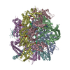
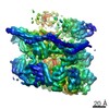
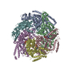
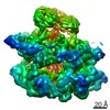
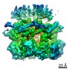
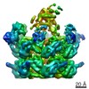
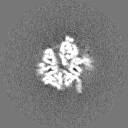
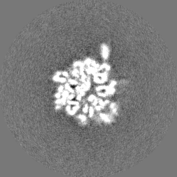
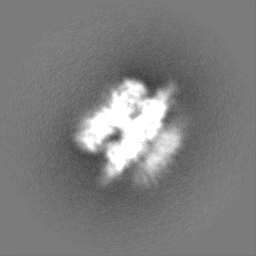
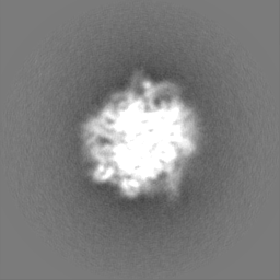
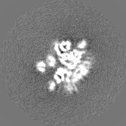
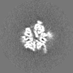
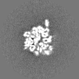
Cryo EM structure of the bacterial disaggregase ClpB (BAP form, DWB mutant), in the ATPgammaS state, bound to the model substrate casein
Map data
Sample
Complex: ClpB homo hexamer bound to the model substract casein
Complex: ClpB homo hexamer bound to the model substract casein
Protein or peptide: ClpB (BAP form, double walker B mutant)
Complex: ClpB homo hexamer bound to the model substract casein
Protein or peptide: Alpha casein
Function / homology
Function and homology information
endopeptidase Clp complex / ATP-dependent peptidase activity / protein quality control for misfolded or incompletely synthesized proteins / protein unfolding / cellular response to heat / response to heat / protein refolding / response to oxidative stress / ATP hydrolysis activity / proteolysis ...endopeptidase Clp complex / ATP-dependent peptidase activity / protein quality control for misfolded or incompletely synthesized proteins / protein unfolding / cellular response to heat / response to heat / protein refolding / response to oxidative stress / ATP hydrolysis activity / proteolysis / ATP binding / membrane / identical protein binding / cytoplasm / cytosol Similarity search - Function
ATP-dependent Clp protease ATP-binding subunit ClpA / Chaperonin ClpB / ClpA/B, conserved site 2 / Chaperonins clpA/B signature 2. / ClpA/B, conserved site 1 / Chaperonins clpA/B signature 1. / ClpA/ClpB, AAA lid domain / AAA lid domain / : / Clp repeat (R) N-terminal domain ...ATP-dependent Clp protease ATP-binding subunit ClpA / Chaperonin ClpB / ClpA/B, conserved site 2 / Chaperonins clpA/B signature 2. / ClpA/B, conserved site 1 / Chaperonins clpA/B signature 1. / ClpA/ClpB, AAA lid domain / AAA lid domain / : / Clp repeat (R) N-terminal domain / Clp repeat (R) domain profile. / Clp, repeat (R) domain / Clp, N-terminal domain superfamily / ClpA/B family / Clp ATPase, C-terminal / C-terminal, D2-small domain, of ClpB protein / C-terminal, D2-small domain, of ClpB protein / AAA domain (Cdc48 subfamily) / ATPase family associated with various cellular activities (AAA) / ATPase, AAA-type, core / ATPases associated with a variety of cellular activities / AAA+ ATPase domain / P-loop containing nucleoside triphosphate hydrolase Similarity search - Domain/homology
Biotechnology and Biological Sciences Research Council
BB/L014211/1
United Kingdom
Hartmut Hoffmann-Berling International Graduate School of Molecular and Cellular Biology
Germany
Knut and Alice Wallenberg Foundation and Family Erling Persson Foundation
Sweden
German Research Foundation
MO 970/4-2
Germany
German Research Foundation
BB617/17-2
Germany
Citation
Journal: Sci Adv / Year: 2017 Title: Structural pathway of regulated substrate transfer and threading through an Hsp100 disaggregase. Authors: Célia Deville / Marta Carroni / Kamila B Franke / Maya Topf / Bernd Bukau / Axel Mogk / Helen R Saibil / Abstract: Refolding aggregated proteins is essential in combating cellular proteotoxic stress. Together with Hsp70, Hsp100 chaperones, including ClpB, form a powerful disaggregation machine that threads ...Refolding aggregated proteins is essential in combating cellular proteotoxic stress. Together with Hsp70, Hsp100 chaperones, including ClpB, form a powerful disaggregation machine that threads aggregated polypeptides through the central pore of tandem adenosine triphosphatase (ATPase) rings. To visualize protein disaggregation, we determined cryo-electron microscopy structures of inactive and substrate-bound ClpB in the presence of adenosine 5'--(3-thiotriphosphate), revealing closed AAA+ rings with a pronounced seam. In the substrate-free state, a marked gradient of resolution, likely corresponding to mobility, spans across the AAA+ rings with a dynamic hotspot at the seam. On the seam side, the coiled-coil regulatory domains are locked in a horizontal, inactive orientation. On the opposite side, the regulatory domains are accessible for Hsp70 binding, substrate targeting, and activation. In the presence of the model substrate casein, the polypeptide threads through the entire pore channel and increased nucleotide occupancy correlates with higher ATPase activity. Substrate-induced domain displacements indicate a pathway of regulated substrate transfer from Hsp70 to the ClpB pore, inside which a spiral of loops contacts the substrate. The seam pore loops undergo marked displacements, along with ordering of the regulatory domains. These asymmetric movements suggest a mechanism for ATPase activation and substrate threading during disaggregation.
History
Deposition
Jun 22, 2017
-
Header (metadata) release
Aug 16, 2017
-
Map release
Aug 16, 2017
-
Update
Nov 6, 2019
-
Current status
Nov 6, 2019
Processing site: PDBe / Status: Released
-
Structure visualization
Movie
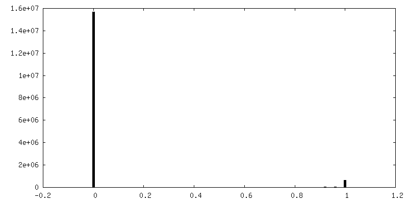
Surface view with section colored by density value
In the structure databanks used in Yorodumi, some data are registered as the other names, "COVID-19 virus" and "2019-nCoV". Here are the details of the virus and the list of structure data.
Jan 31, 2019. EMDB accession codes are about to change! (news from PDBe EMDB page)
EMDB accession codes are about to change! (news from PDBe EMDB page)
The allocation of 4 digits for EMDB accession codes will soon come to an end. Whilst these codes will remain in use, new EMDB accession codes will include an additional digit and will expand incrementally as the available range of codes is exhausted. The current 4-digit format prefixed with “EMD-” (i.e. EMD-XXXX) will advance to a 5-digit format (i.e. EMD-XXXXX), and so on. It is currently estimated that the 4-digit codes will be depleted around Spring 2019, at which point the 5-digit format will come into force.
The EM Navigator/Yorodumi systems omit the EMD- prefix.
Related info.:Q: What is EMD? / ID/Accession-code notation in Yorodumi/EM Navigator
Yorodumi is a browser for structure data from EMDB, PDB, SASBDB, etc.
This page is also the successor to EM Navigator detail page, and also detail information page/front-end page for Omokage search.
The word "yorodu" (or yorozu) is an old Japanese word meaning "ten thousand". "mi" (miru) is to see.
Related info.:EMDB / PDB / SASBDB / Comparison of 3 databanks / Yorodumi Search / Aug 31, 2016. New EM Navigator & Yorodumi / Yorodumi Papers / Jmol/JSmol / Function and homology information / Changes in new EM Navigator and Yorodumi
 Movie
Movie Controller

 Yorodumi
 Open data
 Basic information
 Map data
 Sample
 Function and homology information

 Authors
 United Kingdom,
  Germany,
  Sweden, 9 items
  Citation
 Structure visualization
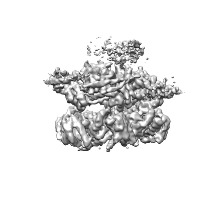
 Downloads & links
 emd_3776.png
 http://ftp.pdbj.org/pub/emdb/structures/EMD-3776


 Z (Sec.)
 Y (Row.)
 X (Col.)


 Sample components
 Processing
 Electron microscopy
 FIELD EMISSION GUN

