Movie
Movie Controller
 Structure viewers
 About Yorodumi Papers


+Search query
-Structure paper
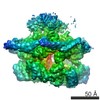
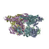
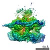
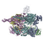
| Title | Structural pathway of regulated substrate transfer and threading through an Hsp100 disaggregase. |
|---|---|
| Journal, issue, pages | Sci Adv, Vol. 3, Issue 8, Page e1701726, Year 2017 |
| Publish date | Aug 4, 2017 |
 Authors Authors | Célia Deville / Marta Carroni / Kamila B Franke / Maya Topf / Bernd Bukau / Axel Mogk / Helen R Saibil /   |
| PubMed Abstract | Refolding aggregated proteins is essential in combating cellular proteotoxic stress. Together with Hsp70, Hsp100 chaperones, including ClpB, form a powerful disaggregation machine that threads ...Refolding aggregated proteins is essential in combating cellular proteotoxic stress. Together with Hsp70, Hsp100 chaperones, including ClpB, form a powerful disaggregation machine that threads aggregated polypeptides through the central pore of tandem adenosine triphosphatase (ATPase) rings. To visualize protein disaggregation, we determined cryo-electron microscopy structures of inactive and substrate-bound ClpB in the presence of adenosine 5'--(3-thiotriphosphate), revealing closed AAA+ rings with a pronounced seam. In the substrate-free state, a marked gradient of resolution, likely corresponding to mobility, spans across the AAA+ rings with a dynamic hotspot at the seam. On the seam side, the coiled-coil regulatory domains are locked in a horizontal, inactive orientation. On the opposite side, the regulatory domains are accessible for Hsp70 binding, substrate targeting, and activation. In the presence of the model substrate casein, the polypeptide threads through the entire pore channel and increased nucleotide occupancy correlates with higher ATPase activity. Substrate-induced domain displacements indicate a pathway of regulated substrate transfer from Hsp70 to the ClpB pore, inside which a spiral of loops contacts the substrate. The seam pore loops undergo marked displacements, along with ordering of the regulatory domains. These asymmetric movements suggest a mechanism for ATPase activation and substrate threading during disaggregation. |
 External links External links | Sci Adv / PubMed:28798962 / PubMed Central |
| Methods | EM (single particle) |
| Resolution | 4.5 - 4.6 Å |
| Structure data | EMDB-3776: Cryo EM structure of the bacterial disaggregase ClpB (BAP form, DWB mutant), in the ATPgammaS state, bound to the model substrate casein |
| Chemicals |  ChemComp-AGS: |
| Source |
|
 Keywords Keywords | CHAPERONE / disaggregase / ClpB / AAA |