 Movie
Movie Controller


+ Open data

- Basic information
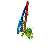
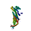
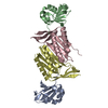
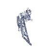
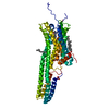
| Entry | Database: PDB / ID: 5oen | ||||||
|---|---|---|---|---|---|---|---|
| Title | Crystal Structure of STAT2 in complex with IRF9 | ||||||
 Components Components |
| ||||||
 Keywords Keywords | TRANSCRIPTION / STAT2 / IRF9 | ||||||
| Function / homology |  Function and homology information Function and homology informationlymphocyte activation involved in immune response / ISGF3 complex / Interferon alpha/beta signaling / lymphocyte differentiation / dendritic cell differentiation / myeloid cell differentiation / defense response to protozoan / immune system process / cytokine-mediated signaling pathway / sequence-specific double-stranded DNA binding ...lymphocyte activation involved in immune response / ISGF3 complex / Interferon alpha/beta signaling / lymphocyte differentiation / dendritic cell differentiation / myeloid cell differentiation / defense response to protozoan / immune system process / cytokine-mediated signaling pathway / sequence-specific double-stranded DNA binding / DNA-binding transcription factor activity, RNA polymerase II-specific / RNA polymerase II cis-regulatory region sequence-specific DNA binding / DNA-binding transcription factor activity / regulation of transcription by RNA polymerase II / positive regulation of transcription by RNA polymerase II / DNA binding / nucleus / cytoplasm / cytosol Similarity search - Function | ||||||
| Biological species |  | ||||||
| Method |  X-RAY DIFFRACTION / SYNCHROTRON / MOLECULAR REPLACEMENT / Resolution: 2.919 Å X-RAY DIFFRACTION / SYNCHROTRON / MOLECULAR REPLACEMENT / Resolution: 2.919 Å | ||||||
 Authors Authors | Rengachari, S. / Panne, D. | ||||||
 Citation Citation | Journal: Proc. Natl. Acad. Sci. U.S.A. / Year: 2018 Title: Structural basis of STAT2 recognition by IRF9 reveals molecular insights into ISGF3 function. Authors: Rengachari, S. / Groiss, S. / Devos, J.M. / Caron, E. / Grandvaux, N. / Panne, D. | ||||||
| History |
|
- Structure visualization
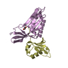
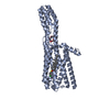
| Structure viewer | Molecule: MolmilJmol/JSmol |
|---|
- Downloads & links
-Download
| PDBx/mmCIF format | 5oen.cif.gz | 136.4 KB | Display | PDBx/mmCIF format |
|---|---|---|---|---|
| PDB format | pdb5oen.ent.gz | 109.2 KB | Display | PDB format |
| PDBx/mmJSON format | 5oen.json.gz | Tree view | PDBx/mmJSON format | |
| Others |  Other downloads Other downloads |
-Validation report
| Arichive directory | https://data.pdbj.org/pub/pdb/validation_reports/oe/5oenftp://data.pdbj.org/pub/pdb/validation_reports/oe/5oen | HTTPS FTP |
|---|
-Related structure data
| Related structure data |  5oemC 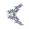 1bf5S S: Starting model for refinement C: citing same article ( |
|---|---|
| Similar structure data |

-Links
 PDBj
PDBj


- Assembly
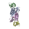
| Deposited unit | 
| ||||||||
|---|---|---|---|---|---|---|---|---|---|
| 1 |
| ||||||||
| Unit cell |
|
-Components
| #1: Protein | Mass: 19100.721 Da / Num. of mol.: 1 / Fragment: UNP residues 206-376 Source method: isolated from a genetically manipulated source Source: (gene. exp.) Production host:  References: UniProt: Q61179 |
|---|---|
| #2: Protein | Mass: 20238.412 Da / Num. of mol.: 1 / Fragment: UNP residues 141-315 Source method: isolated from a genetically manipulated source Source: (gene. exp.) Production host: References: UniProt: Q3UDU1 |
| #3: Water | ChemComp-HOH /  Mass: 18.015 Da / Num. of mol.: 19 / Source method: isolated from a natural source / Formula: H2O Mass: 18.015 Da / Num. of mol.: 19 / Source method: isolated from a natural source / Formula: H2O |
-Experimental details
-Experiment
| Experiment | Method: X-RAY DIFFRACTION / Number of used crystals: 1 |
|---|
- Sample preparation
| Crystal | Density Matthews: 2.46 Å3/Da / Density % sol: 49.92 % |
|---|---|
| Crystal grow | Temperature: 277.15 K / Method: vapor diffusion, hanging drop / Details: 0.2M Potassium formate and 20% PEG3350 |
-Data collection
| Diffraction | Mean temperature: 100 K |
|---|---|
| Diffraction source | Source: SYNCHROTRON / Site: ESRF  / Beamline: ID23-1 / Wavelength: 0.972 Å / Beamline: ID23-1 / Wavelength: 0.972 Å |
| Detector | Type: ADSC QUANTUM 315r / Detector: CCD / Date: Feb 9, 2017 |
| Radiation | Protocol: SINGLE WAVELENGTH / Monochromatic (M) / Laue (L): M / Scattering type: x-ray |
| Radiation wavelength | Wavelength: 0.972 Å / Relative weight: 1 |
| Reflection | Resolution: 2.919→39.389 Å / Num. obs: 7970 / % possible obs: 96 % / Redundancy: 2.8 % / Rmerge(I) obs: 0.194 / Net I/σ(I): 5.9 |
| Reflection shell | Resolution: 2.919→2.93 Å / Redundancy: 2.8 % / Rmerge(I) obs: 0.898 / % possible all: 98.7 |
- Processing
| Software |
| ||||||||||||||||||||||||||||||||||||||||||
|---|---|---|---|---|---|---|---|---|---|---|---|---|---|---|---|---|---|---|---|---|---|---|---|---|---|---|---|---|---|---|---|---|---|---|---|---|---|---|---|---|---|---|---|
| Refinement | Method to determine structure: MOLECULAR REPLACEMENT Starting model: 1BF5 Resolution: 2.919→32.12 Å / SU ML: 0.46 / Cross valid method: THROUGHOUT / σ(F): 1.35 / Phase error: 32.95 / Stereochemistry target values: ML
| ||||||||||||||||||||||||||||||||||||||||||
| Solvent computation | Shrinkage radii: 0.9 Å / VDW probe radii: 1.11 Å / Solvent model: FLAT BULK SOLVENT MODEL | ||||||||||||||||||||||||||||||||||||||||||
| Displacement parameters | Biso mean: 42.46 Å2 | ||||||||||||||||||||||||||||||||||||||||||
| Refinement step | Cycle: LAST / Resolution: 2.919→32.12 Å
| ||||||||||||||||||||||||||||||||||||||||||
| Refine LS restraints |
| ||||||||||||||||||||||||||||||||||||||||||
| LS refinement shell |
|
