 Movie
Movie Controller

[English] 日本語
 Yorodumi
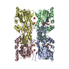
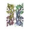
- PDB-5o4h: HcgC from Methanococcus maripaludis cocrystallized with SAM and p... -
+ Open data

- Basic information
| Entry | Database: PDB / ID: 5o4h | ||||||
|---|---|---|---|---|---|---|---|
| Title | HcgC from Methanococcus maripaludis cocrystallized with SAM and pyridinol | ||||||
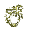
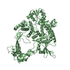
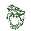
 Components Components | HcgC | ||||||
 Keywords Keywords | TRANSFERASE / methyltransferases / biosynthesis / protein structures / enzyme catalysis / mutagenesis / [Fe]-hydrogenase / pyridinol / Hmd | ||||||
| Function / homology | FeGP cofactor biosynthesis protein, methyltransferase HcgC / FeGP cofactor biosynthesis protein, methyltransferase HcgC / ACETATE ION / S-ADENOSYL-L-HOMOCYSTEINE / Uncharacterized protein Function and homology information Function and homology information | ||||||
| Biological species |  Methanococcus maripaludis S2 (archaea) Methanococcus maripaludis S2 (archaea) | ||||||
| Method |  X-RAY DIFFRACTION / SYNCHROTRON / MOLECULAR REPLACEMENT / Resolution: 1.75 Å X-RAY DIFFRACTION / SYNCHROTRON / MOLECULAR REPLACEMENT / Resolution: 1.75 Å | ||||||
 Authors Authors | Wagner, T. / Bai, L. / Xu, T. / Hu, X. / Ermler, U. / Shima, S. | ||||||
| Funding support |  Germany, 1items Germany, 1items
| ||||||
 Citation Citation | Journal: Angew. Chem. Int. Ed. Engl. / Year: 2017 Title: A Water-Bridged H-Bonding Network Contributes to the Catalysis of the SAM-Dependent C-Methyltransferase HcgC. Authors: Bai, L. / Wagner, T. / Xu, T. / Hu, X. / Ermler, U. / Shima, S. | ||||||
| History |
|
- Structure visualization
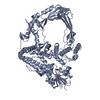
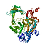
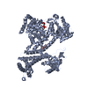
| Structure viewer | Molecule: MolmilJmol/JSmol |
|---|
- Downloads & links
-Download
| PDBx/mmCIF format | 5o4h.cif.gz | 444.5 KB | Display | PDBx/mmCIF format |
|---|---|---|---|---|
| PDB format | pdb5o4h.ent.gz | 362 KB | Display | PDB format |
| PDBx/mmJSON format | 5o4h.json.gz | Tree view | PDBx/mmJSON format | |
| Others |  Other downloads Other downloads |
-Validation report
| Arichive directory | https://data.pdbj.org/pub/pdb/validation_reports/o4/5o4hftp://data.pdbj.org/pub/pdb/validation_reports/o4/5o4h | HTTPS FTP |
|---|
-Related structure data
| Related structure data |  5o4jC 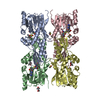 5o4mC  5o4nC  5d4vS S: Starting model for refinement C: citing same article ( |
|---|---|
| Similar structure data |

-Links
 PDBj
PDBj- Assembly
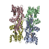
| Deposited unit | 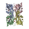
| ||||||||
|---|---|---|---|---|---|---|---|---|---|
| 1 |
| ||||||||
| Unit cell |
|
-Components
-Protein , 1 types, 4 molecules ABCD
| #1: Protein | Mass: 30942.623 Da / Num. of mol.: 4 Source method: isolated from a genetically manipulated source Details: A his-tagged has been placed in the C-terminal of the construct Source: (gene. exp.) Methanococcus maripaludis S2 (archaea) / Cell line: / / Gene: MMP1498 / Production host:  |
|---|
-Non-polymers , 6 types, 718 molecules 


| #2: Chemical | ChemComp-SAH /  Mass: 384.411 Da / Num. of mol.: 4 / Source method: obtained synthetically / Formula: C14H20N6O5S Mass: 384.411 Da / Num. of mol.: 4 / Source method: obtained synthetically / Formula: C14H20N6O5S#3: Chemical | ChemComp-SO4 /  Mass: 96.063 Da / Num. of mol.: 5 / Source method: obtained synthetically / Formula: SO4 Mass: 96.063 Da / Num. of mol.: 5 / Source method: obtained synthetically / Formula: SO4#4: Chemical | ChemComp-ACT /  Mass: 59.044 Da / Num. of mol.: 5 / Source method: obtained synthetically / Formula: C2H3O2 Mass: 59.044 Da / Num. of mol.: 5 / Source method: obtained synthetically / Formula: C2H3O2#5: Chemical | ChemComp-PE4 /  Mass: 354.436 Da / Num. of mol.: 4 / Source method: obtained synthetically / Formula: C16H34O8 / Comment: precipitant*YM Mass: 354.436 Da / Num. of mol.: 4 / Source method: obtained synthetically / Formula: C16H34O8 / Comment: precipitant*YM#6: Chemical | ChemComp-NA / |  Mass: 22.990 Da / Num. of mol.: 1 / Source method: obtained synthetically / Formula: Na Mass: 22.990 Da / Num. of mol.: 1 / Source method: obtained synthetically / Formula: Na#7: Water | ChemComp-HOH / | Mass: 18.015 Da / Num. of mol.: 699 / Source method: isolated from a natural source / Formula: H2O |
|---|
-Details
| Has protein modification | N |
|---|
-Experimental details
-Experiment
| Experiment | Method: X-RAY DIFFRACTION / Number of used crystals: 1 |
|---|
- Sample preparation
| Crystal | Density Matthews: 2.17 Å3/Da / Density % sol: 43.4 % / Description: Thick transparent hexagonal brick |
|---|---|
| Crystal grow | Temperature: 281 K / Method: vapor diffusion, sitting drop / pH: 4.5 Details: 5 mg/ml of pure HcgC containing 2 mM SAM and 2 mM pyridinol was used and mixed at a ratio of 1 ul with 1 ul of precipitant composed of 50% v/v PEG 400, 100 mM NaAcetate pH 4.5 and 200 mM LiSO4 |
-Data collection
| Diffraction | Mean temperature: 100 K |
|---|---|
| Diffraction source | Source: SYNCHROTRON / Site: SLS  / Beamline: X10SA / Wavelength: 0.99999 Å / Beamline: X10SA / Wavelength: 0.99999 Å |
| Detector | Type: DECTRIS PILATUS 6M-F / Detector: PIXEL / Date: Sep 11, 2016 |
| Radiation | Protocol: SINGLE WAVELENGTH / Monochromatic (M) / Laue (L): M / Scattering type: x-ray |
| Radiation wavelength | Wavelength: 0.99999 Å / Relative weight: 1 |
| Reflection | Resolution: 1.75→47.85 Å / Num. obs: 106203 / % possible obs: 99.7 % / Redundancy: 4.8 % / Biso Wilson estimate: 29.2 Å2 / CC1/2: 0.997 / Rmerge(I) obs: 0.082 / Rpim(I) all: 0.041 / Net I/σ(I): 9.7 |
| Reflection shell | Resolution: 1.75→1.84 Å / Redundancy: 4.7 % / Rmerge(I) obs: 0.646 / Mean I/σ(I) obs: 2.2 / Num. unique obs: 15490 / CC1/2: 0.421 / Rpim(I) all: 0.331 / % possible all: 100 |
- Processing
| Software |
| ||||||||||||||||||||||||||||||||||||||||||||||||||||||||||||||||||||||||||||||||||||||||||||||||||||||||||||||||||||||||||||||||||||||||||||||||||||||
|---|---|---|---|---|---|---|---|---|---|---|---|---|---|---|---|---|---|---|---|---|---|---|---|---|---|---|---|---|---|---|---|---|---|---|---|---|---|---|---|---|---|---|---|---|---|---|---|---|---|---|---|---|---|---|---|---|---|---|---|---|---|---|---|---|---|---|---|---|---|---|---|---|---|---|---|---|---|---|---|---|---|---|---|---|---|---|---|---|---|---|---|---|---|---|---|---|---|---|---|---|---|---|---|---|---|---|---|---|---|---|---|---|---|---|---|---|---|---|---|---|---|---|---|---|---|---|---|---|---|---|---|---|---|---|---|---|---|---|---|---|---|---|---|---|---|---|---|---|---|---|---|
| Refinement | Method to determine structure: MOLECULAR REPLACEMENT Starting model: 5D4V Resolution: 1.75→47.85 Å / Cor.coef. Fo:Fc: 0.9603 / Cor.coef. Fo:Fc free: 0.9512 / SU R Cruickshank DPI: 0.15 / Cross valid method: THROUGHOUT / σ(F): 0 / SU R Blow DPI: 0.105 / SU Rfree Blow DPI: 0.098 / SU Rfree Cruickshank DPI: 0.099
| ||||||||||||||||||||||||||||||||||||||||||||||||||||||||||||||||||||||||||||||||||||||||||||||||||||||||||||||||||||||||||||||||||||||||||||||||||||||
| Displacement parameters | Biso mean: 37.97 Å2
| ||||||||||||||||||||||||||||||||||||||||||||||||||||||||||||||||||||||||||||||||||||||||||||||||||||||||||||||||||||||||||||||||||||||||||||||||||||||
| Refine analyze | Luzzati coordinate error obs: 0.215 Å | ||||||||||||||||||||||||||||||||||||||||||||||||||||||||||||||||||||||||||||||||||||||||||||||||||||||||||||||||||||||||||||||||||||||||||||||||||||||
| Refinement step | Cycle: 1 / Resolution: 1.75→47.85 Å
| ||||||||||||||||||||||||||||||||||||||||||||||||||||||||||||||||||||||||||||||||||||||||||||||||||||||||||||||||||||||||||||||||||||||||||||||||||||||
| Refine LS restraints |
| ||||||||||||||||||||||||||||||||||||||||||||||||||||||||||||||||||||||||||||||||||||||||||||||||||||||||||||||||||||||||||||||||||||||||||||||||||||||
| LS refinement shell | Resolution: 1.75→1.79 Å / Total num. of bins used: 20
| ||||||||||||||||||||||||||||||||||||||||||||||||||||||||||||||||||||||||||||||||||||||||||||||||||||||||||||||||||||||||||||||||||||||||||||||||||||||
| Refinement TLS params. | Method: refined / Refine-ID: X-RAY DIFFRACTION
| ||||||||||||||||||||||||||||||||||||||||||||||||||||||||||||||||||||||||||||||||||||||||||||||||||||||||||||||||||||||||||||||||||||||||||||||||||||||
| Refinement TLS group |
|
