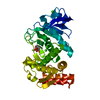
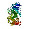
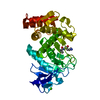
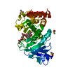
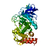
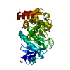
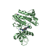
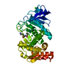
Entry Database : PDB / ID : 5nxdTitle LIM Domain Kinase 2 (LIMK2) In Complex With TH-300 LIM domain kinase 2 Keywords / Function / homology Function Domain/homology Component
/ / / / / / / / / / / / / / / / / / / / / / / / / / / / / / / / / / / / / / / / / / / / / / / / / / / / / / / / / / / / / / / / / Biological species Homo sapiens (human)Method / / / Resolution : 1.9 Å Authors Mathea, S. / Salah, E. / Hanke, T. / Newman, J.A. / Oerum, S. / Wang, D. / Burgess-Brown, N. / von Delft, F. / Arrowsmith, C.H. / Edwards, A.M. ...Mathea, S. / Salah, E. / Hanke, T. / Newman, J.A. / Oerum, S. / Wang, D. / Burgess-Brown, N. / von Delft, F. / Arrowsmith, C.H. / Edwards, A.M. / Bountra, C. / Bullock, A.N. / Knapp, S. Journal : To Be Published Title : LIM Domain Kinase 2 (LIMK2)In Complex With TH-300Authors : Mathea, S. / Salah, E. / Hanke, T. / Newman, J.A. / Oerum, S. / Wang, D. / Burgess-Brown, N. / von Delft, F. / Arrowsmith, C.H. / Edwards, A.M. / Bountra, C. / Bullock, A.N. / Knapp, S. History Deposition May 10, 2017 Deposition site / Processing site Revision 1.0 May 24, 2017 Provider / Type Revision 1.1 Jan 17, 2024 Group / Database references / Refinement descriptionCategory chem_comp_atom / chem_comp_bond ... chem_comp_atom / chem_comp_bond / database_2 / pdbx_initial_refinement_model Item / _database_2.pdbx_database_accession
Show all Show less
 Movie
Movie Controller


 Open data
 Basic information
 Components
 Keywords
 Function and homology information
 Homo sapiens (human)
 X-RAY DIFFRACTION /
  Authors
 Citation
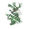
 Structure visualization
 Downloads & links
 Other downloads
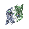


 PDBj
PDBj


 Assembly
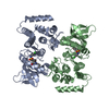


 Spodoptera frugiperda (fall armyworm)
 Mass: 442.958 Da / Num. of mol.: 2 / Source method: obtained synthetically / Formula: C23H23ClN2O3S
 Mass: 62.068 Da / Num. of mol.: 2 / Source method: obtained synthetically / Formula: C2H6O2
 Mass: 18.015 Da / Num. of mol.: 133 / Source method: isolated from a natural source / Formula: H2O
 Sample preparation
 / Beamline: I02 / Wavelength: 0.9795 Å
 Processing