 Movie
Movie Controller

[English] 日本語
 Yorodumi
- PDB-5tak: Conformational Sampling Differences across the Arrhenius Plot Bip... -
+ Open data

- Basic information
| Entry | Database: PDB / ID: 5tak | ||||||
|---|---|---|---|---|---|---|---|
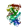
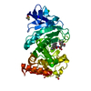
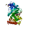
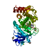
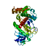
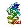
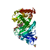
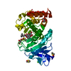
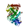
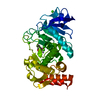
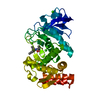
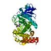
| Title | Conformational Sampling Differences across the Arrhenius Plot Biphasic Break Point at Ambient Temperature in the Enzyme Thermolysin | ||||||
 Components Components | Thermolysin | ||||||
 Keywords Keywords | HYDROLASE / conformational sampling / room temperature / thermolysin / Arrhenius break | ||||||
| Function / homology |  Function and homology information Function and homology informationthermolysin / metalloendopeptidase activity / proteolysis / extracellular region / metal ion binding Similarity search - Function | ||||||
| Biological species |  | ||||||
| Method |  X-RAY DIFFRACTION / MOLECULAR REPLACEMENT / Resolution: 2.003 Å X-RAY DIFFRACTION / MOLECULAR REPLACEMENT / Resolution: 2.003 Å | ||||||
 Authors Authors | Dong, M. / Bahnson, B.J. | ||||||
 Citation Citation | Journal: To Be Published Title: Conformational Sampling Differences across the Arrhenius Plot Biphasic Break Point at Ambient Temperature in the Enzyme Thermolysin Authors: Dong, M. / Bahnson, B.J. | ||||||
| History |
|
- Structure visualization
| Structure viewer | Molecule: MolmilJmol/JSmol |
|---|
- Downloads & links
-Download
| PDBx/mmCIF format | 5tak.cif.gz | 88.7 KB | Display | PDBx/mmCIF format |
|---|---|---|---|---|
| PDB format | pdb5tak.ent.gz | 64.5 KB | Display | PDB format |
| PDBx/mmJSON format | 5tak.json.gz | Tree view | PDBx/mmJSON format | |
| Others |  Other downloads Other downloads |
-Validation report
| Arichive directory | https://data.pdbj.org/pub/pdb/validation_reports/ta/5takftp://data.pdbj.org/pub/pdb/validation_reports/ta/5tak | HTTPS FTP |
|---|
-Related structure data
| Related structure data | 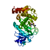 5t9iC  5t9kC 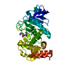 5t9qC 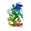 5tacC  5tadC 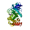 5taeC  5taiC  5tajC  3dnzS C: citing same article ( S: Starting model for refinement |
|---|---|
| Similar structure data |


-Links
 PDBj
PDBj
- Assembly
| Deposited unit | 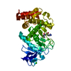
| ||||||||
|---|---|---|---|---|---|---|---|---|---|
| 1 |
| ||||||||
| Unit cell |
| ||||||||
| Components on special symmetry positions |
|
-Components
-Protein , 1 types, 1 molecules A
| #1: Protein | Mass: 34360.336 Da / Num. of mol.: 1 / Fragment: UNP residues 233-548 Source method: isolated from a genetically manipulated source Source: (gene. exp.) Gene: npr / Production host: |
|---|
-Non-polymers , 5 types, 370 molecules 


| #2: Chemical | ChemComp-CA /  Mass: 40.078 Da / Num. of mol.: 4 / Source method: isolated from a natural source / Formula: Ca Mass: 40.078 Da / Num. of mol.: 4 / Source method: isolated from a natural source / Formula: Ca#3: Chemical | ChemComp-ZN / |  Mass: 65.409 Da / Num. of mol.: 1 / Source method: obtained synthetically / Formula: Zn Mass: 65.409 Da / Num. of mol.: 1 / Source method: obtained synthetically / Formula: Zn#4: Chemical | ChemComp-VAL / |  Type: L-peptide linking / Mass: 117.146 Da / Num. of mol.: 1 / Source method: obtained synthetically / Formula: C5H11NO2 Type: L-peptide linking / Mass: 117.146 Da / Num. of mol.: 1 / Source method: obtained synthetically / Formula: C5H11NO2#5: Chemical | ChemComp-LYS / |  Type: L-peptide linking / Mass: 147.195 Da / Num. of mol.: 1 / Source method: obtained synthetically / Formula: C6H15N2O2 Type: L-peptide linking / Mass: 147.195 Da / Num. of mol.: 1 / Source method: obtained synthetically / Formula: C6H15N2O2#6: Water | ChemComp-HOH / | Mass: 18.015 Da / Num. of mol.: 363 / Source method: isolated from a natural source / Formula: H2O |
|---|
-Experimental details
-Experiment
| Experiment | Method: X-RAY DIFFRACTION / Number of used crystals: 1 |
|---|
- Sample preparation
| Crystal | Density Matthews: 2.36 Å3/Da / Density % sol: 47.93 % |
|---|---|
| Crystal grow | Temperature: 293 K / Method: vapor diffusion, sitting drop / pH: 8.3 Details: 1.4 M (NH 4 ) 2 SO 4 , 12% glycerol, 100 mM Tris buffer, pH 8.3 |
-Data collection
| Diffraction | Mean temperature: 93 K |
|---|---|
| Diffraction source | Source: ROTATING ANODE / Type: RIGAKU RUH3R / Wavelength: 1.54178 Å |
| Detector | Type: RIGAKU RAXIS IV / Detector: IMAGE PLATE / Date: May 1, 2012 |
| Radiation | Protocol: SINGLE WAVELENGTH / Monochromatic (M) / Laue (L): M / Scattering type: x-ray |
| Radiation wavelength | Wavelength: 1.54178 Å / Relative weight: 1 |
| Reflection | Resolution: 2→50 Å / Num. obs: 22165 / % possible obs: 99 % / Redundancy: 17.6 % / Net I/σ(I): 22.3 |
- Processing
| Software |
| |||||||||||||||||||||||||||||||||||||||||||||||||||||||||||||||
|---|---|---|---|---|---|---|---|---|---|---|---|---|---|---|---|---|---|---|---|---|---|---|---|---|---|---|---|---|---|---|---|---|---|---|---|---|---|---|---|---|---|---|---|---|---|---|---|---|---|---|---|---|---|---|---|---|---|---|---|---|---|---|---|---|
| Refinement | Method to determine structure: MOLECULAR REPLACEMENT Starting model: 3DNZ Resolution: 2.003→38.506 Å / SU ML: 0.31 / Cross valid method: FREE R-VALUE / σ(F): 1.33 / Phase error: 24.6
| |||||||||||||||||||||||||||||||||||||||||||||||||||||||||||||||
| Solvent computation | Shrinkage radii: 0.98 Å / VDW probe radii: 1.2 Å / Bsol: 57.11 Å2 / ksol: 0.367 e/Å3 | |||||||||||||||||||||||||||||||||||||||||||||||||||||||||||||||
| Displacement parameters |
| |||||||||||||||||||||||||||||||||||||||||||||||||||||||||||||||
| Refinement step | Cycle: LAST / Resolution: 2.003→38.506 Å
| |||||||||||||||||||||||||||||||||||||||||||||||||||||||||||||||
| Refine LS restraints |
| |||||||||||||||||||||||||||||||||||||||||||||||||||||||||||||||
| LS refinement shell |
|
