 Movie
Movie Controller

[English] 日本語
 Yorodumi
- PDB-5ns4: Crystal structures of Cy3 cyanine fluorophores stacked onto the e... -
+ Open data

- Basic information
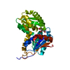
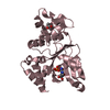
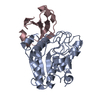
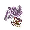
| Entry | Database: PDB / ID: 5ns4 | ||||||
|---|---|---|---|---|---|---|---|
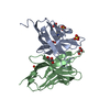
| Title | Crystal structures of Cy3 cyanine fluorophores stacked onto the end of double-stranded RNA | ||||||
 Components Components |
| ||||||
 Keywords Keywords | RNA BINDING PROTEIN / Cy3 / fluorophores / RNA | ||||||
| Function / homology |  Function and homology information Function and homology informationtRNA binding / rRNA binding / structural constituent of ribosome / ribosome / translation / ribonucleoprotein complex Similarity search - Function | ||||||
| Biological species |   Thermus thermophilus (bacteria) Thermus thermophilus (bacteria) | ||||||
| Method |  X-RAY DIFFRACTION / SYNCHROTRON / MOLECULAR REPLACEMENT / Resolution: 2.4 Å X-RAY DIFFRACTION / SYNCHROTRON / MOLECULAR REPLACEMENT / Resolution: 2.4 Å | ||||||
 Authors Authors | Liu, Y.J. / Lilley, D.M.J. | ||||||
| Funding support |  United Kingdom, 1items United Kingdom, 1items
| ||||||
 Citation Citation | Journal: Biophys. J. / Year: 2017 Title: Crystal Structures of Cyanine Fluorophores Stacked onto the End of Double-Stranded RNA. Authors: Liu, Y. / Lilley, D.M.J. | ||||||
| History |
|
- Structure visualization
| Structure viewer | Molecule: MolmilJmol/JSmol |
|---|
- Downloads & links
-Download
| PDBx/mmCIF format | 5ns4.cif.gz | 110.2 KB | Display | PDBx/mmCIF format |
|---|---|---|---|---|
| PDB format | pdb5ns4.ent.gz | 79.4 KB | Display | PDB format |
| PDBx/mmJSON format | 5ns4.json.gz | Tree view | PDBx/mmJSON format | |
| Others |  Other downloads Other downloads |
-Validation report
| Arichive directory | https://data.pdbj.org/pub/pdb/validation_reports/ns/5ns4ftp://data.pdbj.org/pub/pdb/validation_reports/ns/5ns4 | HTTPS FTP |
|---|
-Related structure data
| Related structure data |  5ns3C  1mjiS S: Starting model for refinement C: citing same article ( |
|---|---|
| Similar structure data |


-Links
 PDBj
PDBj


- Assembly
| Deposited unit | 
| ||||||||
|---|---|---|---|---|---|---|---|---|---|
| 1 | 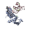
| ||||||||
| 2 | 
| ||||||||
| Unit cell |
|
-Components
| #1: Protein | Mass: 20590.947 Da / Num. of mol.: 2 Source method: isolated from a genetically manipulated source Source: (gene. exp.) Thermus thermophilus (bacteria) / Gene: rplE, rpl5 / Production host: #2: RNA chain | Mass: 10527.345 Da / Num. of mol.: 2 Source method: isolated from a genetically manipulated source Source: (gene. exp.) Thermus thermophilus (bacteria) / Production host: Thermus thermophilus (bacteria)#3: Chemical | ChemComp-MG /   Mass: 24.305 Da / Num. of mol.: 7 / Source method: obtained synthetically / Formula: Mg Mass: 24.305 Da / Num. of mol.: 7 / Source method: obtained synthetically / Formula: Mg#4: Chemical | ChemComp-96T / | 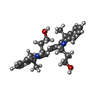  Mass: 445.616 Da / Num. of mol.: 1 Mass: 445.616 Da / Num. of mol.: 1Source method: isolated from a genetically manipulated source Formula: C29H37N2O2 / Source: (gene. exp.) Thermus thermophilus (bacteria) / Production host: Thermus thermophilus (bacteria)#5: Water | ChemComp-HOH / |  Mass: 18.015 Da / Num. of mol.: 72 / Source method: isolated from a natural source / Formula: H2O Mass: 18.015 Da / Num. of mol.: 72 / Source method: isolated from a natural source / Formula: H2O |
|---|
-Experimental details
-Experiment
| Experiment | Method: X-RAY DIFFRACTION / Number of used crystals: 1 |
|---|
- Sample preparation
| Crystal | Density Matthews: 2.35 Å3/Da / Density % sol: 47.73 % |
|---|---|
| Crystal grow | Temperature: 289 K / Method: vapor diffusion, hanging drop Details: 50 mM sodium cacodylate at pH 6.5, 100 mM Mg(CH3COO)2, 50 mM KF, 15% PEG 8000 drop |
-Data collection
| Diffraction | Mean temperature: 93 K |
|---|---|
| Diffraction source | Source: SYNCHROTRON / Site: ESRF  / Beamline: ID23-1 / Wavelength: 0.979601 Å / Beamline: ID23-1 / Wavelength: 0.979601 Å |
| Detector | Type: DECTRIS PILATUS3 S 6M / Detector: PIXEL / Date: Jun 22, 2013 |
| Radiation | Protocol: SINGLE WAVELENGTH / Monochromatic (M) / Laue (L): M / Scattering type: x-ray |
| Radiation wavelength | Wavelength: 0.979601 Å / Relative weight: 1 |
| Reflection | Resolution: 2.4→46.88 Å / Num. obs: 23654 / % possible obs: 100 % / Redundancy: 4.4 % / Biso Wilson estimate: 48.88 Å2 / Rmerge(I) obs: 0.09978 / Rrim(I) all: 0.1141 / Net I/σ(I): 12.48 |
| Reflection shell | Resolution: 2.4→2.486 Å / Redundancy: 4.5 % / Rmerge(I) obs: 0.162 / Mean I/σ(I) obs: 6.26 / Num. unique obs: 2309 / Rrim(I) all: 0.1834 / % possible all: 100 |
- Processing
| Software |
| ||||||||||||||||||||||||||||||||||||||||||||||||||||||||||||||||||||||
|---|---|---|---|---|---|---|---|---|---|---|---|---|---|---|---|---|---|---|---|---|---|---|---|---|---|---|---|---|---|---|---|---|---|---|---|---|---|---|---|---|---|---|---|---|---|---|---|---|---|---|---|---|---|---|---|---|---|---|---|---|---|---|---|---|---|---|---|---|---|---|---|
| Refinement | Method to determine structure: MOLECULAR REPLACEMENT Starting model: 1MJI Resolution: 2.4→46.88 Å / SU ML: 0.34 / Cross valid method: FREE R-VALUE / σ(F): 1.35 / Phase error: 29
| ||||||||||||||||||||||||||||||||||||||||||||||||||||||||||||||||||||||
| Solvent computation | Shrinkage radii: 0.9 Å / VDW probe radii: 1.11 Å | ||||||||||||||||||||||||||||||||||||||||||||||||||||||||||||||||||||||
| Displacement parameters | Biso max: 112.67 Å2 / Biso mean: 37.1008 Å2 / Biso min: 20 Å2 | ||||||||||||||||||||||||||||||||||||||||||||||||||||||||||||||||||||||
| Refinement step | Cycle: final / Resolution: 2.4→46.88 Å
| ||||||||||||||||||||||||||||||||||||||||||||||||||||||||||||||||||||||
| Refine LS restraints |
| ||||||||||||||||||||||||||||||||||||||||||||||||||||||||||||||||||||||
| LS refinement shell | Refine-ID: X-RAY DIFFRACTION / Rfactor Rfree error: 0 / Total num. of bins used: 9
|
