 Movie
Movie Controller

[English] 日本語
 Yorodumi
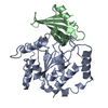
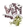
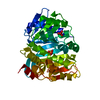
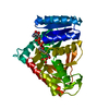
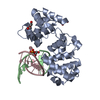
- PDB-2uug: ESCHERICHIA COLI URACIL-DNA GLYCOSYLASE:INHIBITOR COMPLEX WITH H1... -
+ Open data

- Basic information
| Entry | Database: PDB / ID: 2uug | ||||||
|---|---|---|---|---|---|---|---|
| Title | ESCHERICHIA COLI URACIL-DNA GLYCOSYLASE:INHIBITOR COMPLEX WITH H187D MUTANT UDG AND WILD-TYPE UGI | ||||||
 Components Components |
| ||||||
 Keywords Keywords | REPLICATION / HYDROLASE / DNA BASE EXCISION REPAIR / PROTEIN MIMICRY OF DNA / PROTEIN INHIBITOR | ||||||
| Function / homology |  Function and homology information Function and homology informationbase-excision repair, AP site formation via deaminated base removal / uracil-DNA glycosylase / uracil DNA N-glycosylase activity / cytoplasm Similarity search - Function | ||||||
| Biological species |   Bacillus phage PBS2 (virus) Bacillus phage PBS2 (virus) | ||||||
| Method |  X-RAY DIFFRACTION / MOLECULAR REPLACEMENT / Resolution: 2.6 Å X-RAY DIFFRACTION / MOLECULAR REPLACEMENT / Resolution: 2.6 Å | ||||||
 Authors Authors | Putnam, C.D. / Arvai, A.S. / Mol, C.D. / Tainer, J.A. | ||||||
 Citation Citation | Journal: J.Mol.Biol. / Year: 1999 Title: Protein mimicry of DNA from crystal structures of the uracil-DNA glycosylase inhibitor protein and its complex with Escherichia coli uracil-DNA glycosylase Authors: Putnam, C.D. / Shroyer, M.J. / Lundquist, A.J. / Mol, C.D. / Arvai, A.S. / Mosbaugh, D.W. / Tainer, J.A. | ||||||
| History |
|
- Structure visualization
| Structure viewer | Molecule: MolmilJmol/JSmol |
|---|
- Downloads & links
-Download
| PDBx/mmCIF format | 2uug.cif.gz | 128.4 KB | Display | PDBx/mmCIF format |
|---|---|---|---|---|
| PDB format | pdb2uug.ent.gz | 102 KB | Display | PDB format |
| PDBx/mmJSON format | 2uug.json.gz | Tree view | PDBx/mmJSON format | |
| Others |  Other downloads Other downloads |
-Validation report
| Arichive directory | https://data.pdbj.org/pub/pdb/validation_reports/uu/2uugftp://data.pdbj.org/pub/pdb/validation_reports/uu/2uug | HTTPS FTP |
|---|
-Related structure data


-Links
 PDBj
PDBj- Assembly
| Deposited unit | 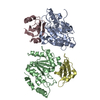
| ||||||||||||
|---|---|---|---|---|---|---|---|---|---|---|---|---|---|
| 1 | 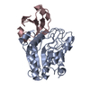
| ||||||||||||
| 2 | 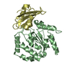
| ||||||||||||
| Unit cell |
| ||||||||||||
| Noncrystallographic symmetry (NCS) | NCS oper:
|
-Components
| #1: Protein | Mass: 25702.123 Da / Num. of mol.: 2 / Mutation: H187D Source method: isolated from a genetically manipulated source Source: (gene. exp.) #2: Protein | Mass: 9482.674 Da / Num. of mol.: 2 Source method: isolated from a genetically manipulated source Source: (gene. exp.) Bacillus phage PBS2 (virus) / Gene: UGI / Plasmid: PZWTAC1 / Cellular location (production host): CYTOPLASM / Gene (production host): TAC / Production host: #3: Water | ChemComp-HOH / |  Mass: 18.015 Da / Num. of mol.: 62 / Source method: isolated from a natural source / Formula: H2O Mass: 18.015 Da / Num. of mol.: 62 / Source method: isolated from a natural source / Formula: H2OSequence details | ENGINEERED | |
|---|
-Experimental details
-Experiment
| Experiment | Method: X-RAY DIFFRACTION / Number of used crystals: 1 |
|---|
- Sample preparation
| Crystal | Density Matthews: 2.1 Å3/Da / Density % sol: 40 % | ||||||||||||||||||||||||||||||
|---|---|---|---|---|---|---|---|---|---|---|---|---|---|---|---|---|---|---|---|---|---|---|---|---|---|---|---|---|---|---|---|
| Crystal grow | pH: 8.2 / Details: pH 8.2 | ||||||||||||||||||||||||||||||
| Components of the solutions |
| ||||||||||||||||||||||||||||||
| Crystal grow | *PLUS Method: vapor diffusion | ||||||||||||||||||||||||||||||
| Components of the solutions | *PLUS
|
-Data collection
| Diffraction | Mean temperature: 150 K |
|---|---|
| Diffraction source | Source: ROTATING ANODE / Type: SIEMENS |
| Detector | Type: MARRESEARCH / Detector: IMAGE PLATE |
| Radiation | Monochromator: GRAPHITE / Protocol: SINGLE WAVELENGTH / Monochromatic (M) / Laue (L): M / Scattering type: x-ray |
| Radiation wavelength | Relative weight: 1 |
| Reflection | Resolution: 2.6→40 Å / Num. obs: 17739 / % possible obs: 99.9 % / Observed criterion σ(I): 0 / Redundancy: 4.6 % / Biso Wilson estimate: 50.9 Å2 / Rsym value: 0.121 / Net I/σ(I): 13.6 |
| Reflection shell | Resolution: 2.6→2.7 Å / Redundancy: 3 % / Mean I/σ(I) obs: 2.9 / Rsym value: 0.457 / % possible all: 99.9 |
| Reflection | *PLUS Num. measured all: 82224 / Rmerge(I) obs: 0.121 |
| Reflection shell | *PLUS % possible obs: 99.9 % / Num. unique obs: 1736 / Rmerge(I) obs: 0.457 |
- Processing
| Software |
| ||||||||||||||||||||||||||||||||||||||||||||||||||||||||||||
|---|---|---|---|---|---|---|---|---|---|---|---|---|---|---|---|---|---|---|---|---|---|---|---|---|---|---|---|---|---|---|---|---|---|---|---|---|---|---|---|---|---|---|---|---|---|---|---|---|---|---|---|---|---|---|---|---|---|---|---|---|---|
| Refinement | Method to determine structure: MOLECULAR REPLACEMENT Starting model: WILD TYPE E. COLI UDG:UGI COMPLEX Resolution: 2.6→20 Å / Data cutoff high absF: 100000 / Data cutoff low absF: 0.1 / Isotropic thermal model: RESTRAINED / Cross valid method: THROUGHOUT / σ(F): 0 Details: PHE A 77 AND PHE B 77 ARE CONSERVED RAMACHANDRAN OUTLIERS IN HUMAN, HSV AND E.COLI UDG
| ||||||||||||||||||||||||||||||||||||||||||||||||||||||||||||
| Displacement parameters | Biso mean: 36.7 Å2
| ||||||||||||||||||||||||||||||||||||||||||||||||||||||||||||
| Refinement step | Cycle: LAST / Resolution: 2.6→20 Å
| ||||||||||||||||||||||||||||||||||||||||||||||||||||||||||||
| Refine LS restraints |
| ||||||||||||||||||||||||||||||||||||||||||||||||||||||||||||
| LS refinement shell | Resolution: 2.6→2.72 Å / Total num. of bins used: 8
| ||||||||||||||||||||||||||||||||||||||||||||||||||||||||||||
| Xplor file |
| ||||||||||||||||||||||||||||||||||||||||||||||||||||||||||||
| Software | *PLUS Name: X-PLOR / Version: 3.851 / Classification: refinement | ||||||||||||||||||||||||||||||||||||||||||||||||||||||||||||
| Refinement | *PLUS Highest resolution: 2.6 Å / Lowest resolution: 20 Å / σ(F): 0 / % reflection Rfree: 10 % / Rfactor obs: 0.178 | ||||||||||||||||||||||||||||||||||||||||||||||||||||||||||||
| Solvent computation | *PLUS | ||||||||||||||||||||||||||||||||||||||||||||||||||||||||||||
| Displacement parameters | *PLUS Biso mean: 36.7 Å2 | ||||||||||||||||||||||||||||||||||||||||||||||||||||||||||||
| Refine LS restraints | *PLUS
| ||||||||||||||||||||||||||||||||||||||||||||||||||||||||||||
| LS refinement shell | *PLUS Highest resolution: 2.6 Å / Rfactor Rfree: 0.323 / % reflection Rfree: 10 % / Rfactor Rwork: 0.286 |
