 Movie
Movie Controller

[English] 日本語
 Yorodumi
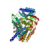
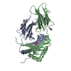
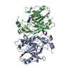
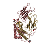
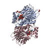
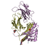
- PDB-5n6m: Structure of the membrane integral lipoprotein N-acyltransferase ... -
+ Open data

- Basic information
| Entry | Database: PDB / ID: 5n6m | ||||||
|---|---|---|---|---|---|---|---|
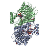
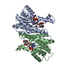
| Title | Structure of the membrane integral lipoprotein N-acyltransferase Lnt from P. aeruginosa | ||||||
 Components Components | Apolipoprotein N-acyltransferase | ||||||
 Keywords Keywords | MEMBRANE PROTEIN / lipoprotein / N-acyltransferase / lipidic cubic phase | ||||||
| Function / homology |  Function and homology information Function and homology informationapolipoprotein N-acyltransferase / N-acyltransferase activity / lipoprotein biosynthetic process / plasma membrane Similarity search - Function | ||||||
| Biological species |   Pseudomonas aeruginosa (bacteria) Pseudomonas aeruginosa (bacteria) | ||||||
| Method |  X-RAY DIFFRACTION / SYNCHROTRON / MOLECULAR REPLACEMENT / Resolution: 3.1 Å X-RAY DIFFRACTION / SYNCHROTRON / MOLECULAR REPLACEMENT / Resolution: 3.1 Å | ||||||
 Authors Authors | Huang, C.-Y. / Boland, C. / Howe, N. / Wiktor, M. / Vogeley, L. / Weichert, D. / Bailey, J. / Olieric, V. / Wang, M. / Caffrey, M. | ||||||
| Funding support |  Ireland, 1items Ireland, 1items
| ||||||
 Citation Citation | Journal: Nat Commun / Year: 2017 Title: Structural insights into the mechanism of the membrane integral N-acyltransferase step in bacterial lipoprotein synthesis. Authors: Wiktor, M. / Weichert, D. / Howe, N. / Huang, C.Y. / Olieric, V. / Boland, C. / Bailey, J. / Vogeley, L. / Stansfeld, P.J. / Buddelmeijer, N. / Wang, M. / Caffrey, M. | ||||||
| History |
|
- Structure visualization
| Structure viewer | Molecule: MolmilJmol/JSmol |
|---|
- Downloads & links
-Download
| PDBx/mmCIF format | 5n6m.cif.gz | 112.7 KB | Display | PDBx/mmCIF format |
|---|---|---|---|---|
| PDB format | pdb5n6m.ent.gz | 84.1 KB | Display | PDB format |
| PDBx/mmJSON format | 5n6m.json.gz | Tree view | PDBx/mmJSON format | |
| Others |  Other downloads Other downloads |
-Validation report
| Summary document | 5n6m_validation.pdf.gz | 1.2 MB | Display | wwPDB validaton report |
|---|---|---|---|---|
| Full document | 5n6m_full_validation.pdf.gz | 1.2 MB | Display | |
| Data in XML | 5n6m_validation.xml.gz | 20.9 KB | Display | |
| Data in CIF | 5n6m_validation.cif.gz | 27.7 KB | Display | |
| Arichive directory | https://data.pdbj.org/pub/pdb/validation_reports/n6/5n6mftp://data.pdbj.org/pub/pdb/validation_reports/n6/5n6m | HTTPS FTP |
-Related structure data
| Related structure data | 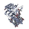 5n6hSC  5n6lC S: Starting model for refinement C: citing same article ( |
|---|---|
| Similar structure data |

-Links
 PDBj
PDBj


- Assembly
| Deposited unit | 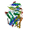
| ||||||||
|---|---|---|---|---|---|---|---|---|---|
| 1 |
| ||||||||
| Unit cell |
|
-Components
| #1: Protein | Mass: 58230.461 Da / Num. of mol.: 1 Source method: isolated from a genetically manipulated source Source: (gene. exp.) Pseudomonas aeruginosa (strain ATCC 15692 / DSM 22644 / CIP 104116 / JCM 14847 / LMG 12228 / 1C / PRS 101 / PAO1) (bacteria)Strain: ATCC 15692 / DSM 22644 / CIP 104116 / JCM 14847 / LMG 12228 / 1C / PRS 101 / PAO1 Gene: lnt, cutE, PA3984 / Cell line (production host): C41 / Production host: Pseudomonas aeruginosa (bacteria)References: UniProt: Q9ZI86, Transferases; Acyltransferases; Transferring groups other than aminoacyl groups | ||||||
|---|---|---|---|---|---|---|---|
| #2: Chemical |   Mass: 92.094 Da / Num. of mol.: 3 / Source method: obtained synthetically / Formula: C3H8O3 Mass: 92.094 Da / Num. of mol.: 3 / Source method: obtained synthetically / Formula: C3H8O3#3: Chemical | ChemComp-FLC / |   Mass: 189.100 Da / Num. of mol.: 1 / Source method: obtained synthetically / Formula: C6H5O7 Mass: 189.100 Da / Num. of mol.: 1 / Source method: obtained synthetically / Formula: C6H5O7#4: Chemical | ChemComp-OLC / (   Mass: 356.540 Da / Num. of mol.: 5 / Source method: obtained synthetically / Formula: C21H40O4 Mass: 356.540 Da / Num. of mol.: 5 / Source method: obtained synthetically / Formula: C21H40O4#5: Water | ChemComp-HOH / |  Mass: 18.015 Da / Num. of mol.: 16 / Source method: isolated from a natural source / Formula: H2O Mass: 18.015 Da / Num. of mol.: 16 / Source method: isolated from a natural source / Formula: H2O |
-Experimental details
-Experiment
| Experiment | Method: X-RAY DIFFRACTION / Number of used crystals: 1 |
|---|
- Sample preparation
| Crystal | Density Matthews: 2.36 Å3/Da / Density % sol: 47.85 % |
|---|---|
| Crystal grow | Temperature: 293 K / Method: lipidic cubic phase Details: 100 mM NaCitrate pH 5.0 30 % (v/v) PEG-500 DME 100 mM sodium acetate |
-Data collection
| Diffraction | Mean temperature: 100 K |
|---|---|
| Diffraction source | Source: SYNCHROTRON / Site: SLS  / Beamline: X06SA / Wavelength: 1.0003 Å / Beamline: X06SA / Wavelength: 1.0003 Å |
| Detector | Type: DECTRIS PILATUS 6M-F / Detector: PIXEL / Date: Jul 4, 2016 |
| Radiation | Monochromator: Si(111) / Protocol: SINGLE WAVELENGTH / Monochromatic (M) / Laue (L): M / Scattering type: x-ray |
| Radiation wavelength | Wavelength: 1.0003 Å / Relative weight: 1 |
| Reflection | Resolution: 3.1→49.5 Å / Num. obs: 10159 / % possible obs: 98.9 % / Redundancy: 5.4 % / Net I/σ(I): 6.48 |
- Processing
| Software |
| ||||||||||||||||||||||||||||||||||||||||||||||||||||||||
|---|---|---|---|---|---|---|---|---|---|---|---|---|---|---|---|---|---|---|---|---|---|---|---|---|---|---|---|---|---|---|---|---|---|---|---|---|---|---|---|---|---|---|---|---|---|---|---|---|---|---|---|---|---|---|---|---|---|
| Refinement | Method to determine structure: MOLECULAR REPLACEMENT Starting model: 5N6H Resolution: 3.1→49.5 Å / SU ML: 0.57 / Cross valid method: FREE R-VALUE / σ(F): 1.35 / Phase error: 29.84
| ||||||||||||||||||||||||||||||||||||||||||||||||||||||||
| Solvent computation | Shrinkage radii: 0.9 Å / VDW probe radii: 1.11 Å | ||||||||||||||||||||||||||||||||||||||||||||||||||||||||
| Refinement step | Cycle: LAST / Resolution: 3.1→49.5 Å
| ||||||||||||||||||||||||||||||||||||||||||||||||||||||||
| Refine LS restraints |
| ||||||||||||||||||||||||||||||||||||||||||||||||||||||||
| LS refinement shell |
|
