 Movie
Movie Controller

[English] 日本語
 Yorodumi
- PDB-5n5u: Structure of p-boronophenylalanyl tRNA synthetase in complex with... -
+ Open data

- Basic information
| Entry | Database: PDB / ID: 5n5u | |||||||||
|---|---|---|---|---|---|---|---|---|---|---|
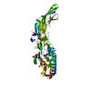
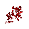
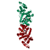
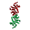
| Title | Structure of p-boronophenylalanyl tRNA synthetase in complex with p-boronophenylalanine and adenosine monophosphate | |||||||||
 Components Components | Tyrosine--tRNA ligase | |||||||||
 Keywords Keywords | LIGASE / aminoacylation / non-natural amino acid / synthetic biology | |||||||||
| Function / homology |  Function and homology information Function and homology informationtyrosyl-tRNA aminoacylation / tyrosine-tRNA ligase / tyrosine-tRNA ligase activity / ATP binding / cytoplasm Similarity search - Function | |||||||||
| Biological species |   Methanocaldococcus jannaschii (archaea) Methanocaldococcus jannaschii (archaea) | |||||||||
| Method |  X-RAY DIFFRACTION / SYNCHROTRON / MOLECULAR REPLACEMENT / Resolution: 1.6 Å X-RAY DIFFRACTION / SYNCHROTRON / MOLECULAR REPLACEMENT / Resolution: 1.6 Å | |||||||||
 Authors Authors | Schiefner, A. / Skerra, A. | |||||||||
 Citation Citation | Journal: Biochemistry / Year: 2018 Title: Structural Basis for the Specific Cotranslational Incorporation of p-Boronophenylalanine into Biosynthetic Proteins. Authors: Schiefner, A. / Nastle, L. / Landgraf, M. / Reichert, A.J. / Skerra, A. | |||||||||
| History |
|
- Structure visualization
| Structure viewer | Molecule: MolmilJmol/JSmol |
|---|
- Downloads & links
-Download
| PDBx/mmCIF format | 5n5u.cif.gz | 147 KB | Display | PDBx/mmCIF format |
|---|---|---|---|---|
| PDB format | pdb5n5u.ent.gz | 115.1 KB | Display | PDB format |
| PDBx/mmJSON format | 5n5u.json.gz | Tree view | PDBx/mmJSON format | |
| Others |  Other downloads Other downloads |
-Validation report
| Arichive directory | https://data.pdbj.org/pub/pdb/validation_reports/n5/5n5uftp://data.pdbj.org/pub/pdb/validation_reports/n5/5n5u | HTTPS FTP |
|---|
-Related structure data
| Related structure data |  5n5vC  4ndaS C: citing same article ( S: Starting model for refinement |
|---|---|
| Similar structure data |


-Links
 PDBj
PDBj


- Assembly
| Deposited unit | 
| ||||||||
|---|---|---|---|---|---|---|---|---|---|
| 1 | 
| ||||||||
| Unit cell |
|
-Components
| #1: Protein | Mass: 35976.734 Da / Num. of mol.: 1 / Mutation: M6L, Y32S, L65A, H70M, D158S, L162E Source method: isolated from a genetically manipulated source Source: (gene. exp.) Methanocaldococcus jannaschii (strain ATCC 43067 / DSM 2661 / JAL-1 / JCM 10045 / NBRC 100440) (archaea)Strain: ATCC 43067 / DSM 2661 / JAL-1 / JCM 10045 / NBRC 100440 Gene: tyrS, MJ0389 / Plasmid: pASK75-BpaRS-His / Production host:  | ||
|---|---|---|---|
| #2: Chemical | ChemComp-AMP /   Mass: 347.221 Da / Num. of mol.: 1 / Source method: obtained synthetically / Formula: C10H14N5O7P / Comment: AMP*YM Mass: 347.221 Da / Num. of mol.: 1 / Source method: obtained synthetically / Formula: C10H14N5O7P / Comment: AMP*YM | ||
| #3: Chemical | ChemComp-7N8 /   Type: L-peptide linking / Mass: 209.007 Da / Num. of mol.: 1 / Source method: obtained synthetically / Formula: C9H12BNO4 Type: L-peptide linking / Mass: 209.007 Da / Num. of mol.: 1 / Source method: obtained synthetically / Formula: C9H12BNO4 | ||
| #4: Chemical |   Mass: 35.453 Da / Num. of mol.: 3 / Source method: obtained synthetically / Formula: Cl Mass: 35.453 Da / Num. of mol.: 3 / Source method: obtained synthetically / Formula: Cl#5: Water | ChemComp-HOH / |  Mass: 18.015 Da / Num. of mol.: 221 / Source method: isolated from a natural source / Formula: H2O Mass: 18.015 Da / Num. of mol.: 221 / Source method: isolated from a natural source / Formula: H2O |
-Experimental details
-Experiment
| Experiment | Method: X-RAY DIFFRACTION / Number of used crystals: 1 |
|---|
- Sample preparation
| Crystal | Density Matthews: 2.32 Å3/Da / Density % sol: 47.03 % |
|---|---|
| Crystal grow | Temperature: 293 K / Method: vapor diffusion, sitting drop / pH: 6.5 Details: 20 % (w/v) PEG 1000, 0.2 M MgCl2, 0.1 M sodium cacodylate |
-Data collection
| Diffraction | Mean temperature: 100 K | ||||||||||||||||||||||||||||||||||||||||||||||||||||||||||||||||||||||||||||||||
|---|---|---|---|---|---|---|---|---|---|---|---|---|---|---|---|---|---|---|---|---|---|---|---|---|---|---|---|---|---|---|---|---|---|---|---|---|---|---|---|---|---|---|---|---|---|---|---|---|---|---|---|---|---|---|---|---|---|---|---|---|---|---|---|---|---|---|---|---|---|---|---|---|---|---|---|---|---|---|---|---|---|
| Diffraction source | Source: SYNCHROTRON / Site: BESSY  / Beamline: 14.3 / Wavelength: 0.8943 Å / Beamline: 14.3 / Wavelength: 0.8943 Å | ||||||||||||||||||||||||||||||||||||||||||||||||||||||||||||||||||||||||||||||||
| Detector | Type: MARMOSAIC 225 mm CCD / Detector: CCD / Date: Jun 29, 2016 | ||||||||||||||||||||||||||||||||||||||||||||||||||||||||||||||||||||||||||||||||
| Radiation | Monochromator: Si111 / Protocol: SINGLE WAVELENGTH / Monochromatic (M) / Laue (L): M / Scattering type: x-ray | ||||||||||||||||||||||||||||||||||||||||||||||||||||||||||||||||||||||||||||||||
| Radiation wavelength | Wavelength: 0.8943 Å / Relative weight: 1 | ||||||||||||||||||||||||||||||||||||||||||||||||||||||||||||||||||||||||||||||||
| Reflection | Resolution: 1.6→30 Å / Num. obs: 43906 / % possible obs: 99.7 % / Observed criterion σ(I): -3 / Redundancy: 7.43 % / Biso Wilson estimate: 31.967 Å2 / CC1/2: 1 / Rmerge(I) obs: 0.043 / Rrim(I) all: 0.046 / Χ2: 1.007 / Net I/σ(I): 24.36 / Num. measured all: 326242 / Scaling rejects: 24 | ||||||||||||||||||||||||||||||||||||||||||||||||||||||||||||||||||||||||||||||||
| Reflection shell | Diffraction-ID: 1
|
- Processing
| Software |
| |||||||||||||||||||||||||||||||||||||||||||||||||||||||||||||||||||||||||||||||||||||||||||||||||||||||||||||||||||||||||||||
|---|---|---|---|---|---|---|---|---|---|---|---|---|---|---|---|---|---|---|---|---|---|---|---|---|---|---|---|---|---|---|---|---|---|---|---|---|---|---|---|---|---|---|---|---|---|---|---|---|---|---|---|---|---|---|---|---|---|---|---|---|---|---|---|---|---|---|---|---|---|---|---|---|---|---|---|---|---|---|---|---|---|---|---|---|---|---|---|---|---|---|---|---|---|---|---|---|---|---|---|---|---|---|---|---|---|---|---|---|---|---|---|---|---|---|---|---|---|---|---|---|---|---|---|---|---|---|
| Refinement | Method to determine structure: MOLECULAR REPLACEMENT Starting model: 4NDA Resolution: 1.6→30 Å / Cor.coef. Fo:Fc: 0.967 / Cor.coef. Fo:Fc free: 0.954 / SU B: 4.119 / SU ML: 0.07 / SU R Cruickshank DPI: 0.0864 / Cross valid method: THROUGHOUT / σ(F): 0 / ESU R: 0.086 / ESU R Free: 0.089 Details: HYDROGENS HAVE BEEN ADDED IN THE RIDING POSITIONS U VALUES : WITH TLS ADDED
| |||||||||||||||||||||||||||||||||||||||||||||||||||||||||||||||||||||||||||||||||||||||||||||||||||||||||||||||||||||||||||||
| Solvent computation | Ion probe radii: 0.8 Å / Shrinkage radii: 0.8 Å / VDW probe radii: 1.2 Å | |||||||||||||||||||||||||||||||||||||||||||||||||||||||||||||||||||||||||||||||||||||||||||||||||||||||||||||||||||||||||||||
| Displacement parameters | Biso max: 116.05 Å2 / Biso mean: 33.232 Å2 / Biso min: 17 Å2
| |||||||||||||||||||||||||||||||||||||||||||||||||||||||||||||||||||||||||||||||||||||||||||||||||||||||||||||||||||||||||||||
| Refinement step | Cycle: final / Resolution: 1.6→30 Å
| |||||||||||||||||||||||||||||||||||||||||||||||||||||||||||||||||||||||||||||||||||||||||||||||||||||||||||||||||||||||||||||
| Refine LS restraints |
| |||||||||||||||||||||||||||||||||||||||||||||||||||||||||||||||||||||||||||||||||||||||||||||||||||||||||||||||||||||||||||||
| LS refinement shell | Resolution: 1.6→1.641 Å / Rfactor Rfree error: 0 / Total num. of bins used: 20
| |||||||||||||||||||||||||||||||||||||||||||||||||||||||||||||||||||||||||||||||||||||||||||||||||||||||||||||||||||||||||||||
| Refinement TLS params. | Method: refined / Refine-ID: X-RAY DIFFRACTION
| |||||||||||||||||||||||||||||||||||||||||||||||||||||||||||||||||||||||||||||||||||||||||||||||||||||||||||||||||||||||||||||
| Refinement TLS group |
|
