- PDB-5mmt: Inward open PepTSt from Streptococcus thermophilus crystallized i... -
+
Open data
ID or keywords:
Loading...
-
Basic information
Entry
Database: PDB / ID: 5mmt
Title
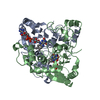
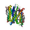
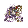
Inward open PepTSt from Streptococcus thermophilus crystallized in space group P3121
Components
Di-or tripeptide:H+ symporter
Keywords
TRANSPORT PROTEIN / Integral membrane protein / Major facilitator superfamily transporter (MFS transporter) / POT PTR or PepT family / proton-coupled peptide transporter
Function / homology
Function and homology information
tripeptide transmembrane transport / tripeptide transmembrane transporter activity / peptide:proton symporter activity / dipeptide transmembrane transporter activity / identical protein binding / plasma membrane Similarity search - Function
: / Dipeptide/tripeptide permease / PTR2 family proton/oligopeptide symporters signature 1. / MFS general substrate transporter like domains / PTR2 family proton/oligopeptide symporters signature 2. / PTR2 family proton/oligopeptide symporter, conserved site / Proton-dependent oligopeptide transporter family / POT family / Growth Hormone; Chain: A; / Major facilitator superfamily domain ...: / Dipeptide/tripeptide permease / PTR2 family proton/oligopeptide symporters signature 1. / MFS general substrate transporter like domains / PTR2 family proton/oligopeptide symporters signature 2. / PTR2 family proton/oligopeptide symporter, conserved site / Proton-dependent oligopeptide transporter family / POT family / Growth Hormone; Chain: A; / Major facilitator superfamily domain / Major facilitator superfamily (MFS) profile. / MFS transporter superfamily / Up-down Bundle / Mainly Alpha Similarity search - Domain/homology
Monochromator: Double crystal / Protocol: SINGLE WAVELENGTH / Monochromatic (M) / Laue (L): M / Scattering type: x-ray
Radiation wavelength
Wavelength: 0.9795 Å / Relative weight: 1
Reflection
Resolution: 3.4→44.85 Å / Num. obs: 11881 / % possible obs: 74.6 % / Redundancy: 5.3 % / Biso Wilson estimate: 96.4 Å2 / CC1/2: 0.999 / Rmerge(I) obs: 0.09 / Rsym value: 0.08 / Net I/σ(I): 13.67
Reflection shell
Resolution: 3.4→3.49 Å / Redundancy: 0.5 % / Rmerge(I) obs: 0.68 / Mean I/σ(I) obs: 3.13 / CC1/2: 0.834 / % possible all: 5.8
-
Processing
Software
Name
Version
Classification
PHENIX
1.9_1692
refinement
XDS
datareduction
XSCALE
datascaling
SHARP
phasing
Refinement
Method to determine structure: MAD / Resolution: 3.4→44.85 Å / SU ML: 0.37 / Cross valid method: FREE R-VALUE / Phase error: 36.6 Details: The data set used for refinement was truncated and anisotropically scaled using the Diffraction Anisotropy Server (https://services.mbi.ucla.edu/anisoscale/). Cut-off values for truncation: ...Details: The data set used for refinement was truncated and anisotropically scaled using the Diffraction Anisotropy Server (https://services.mbi.ucla.edu/anisoscale/). Cut-off values for truncation: 3.9 A in the a-direction, 3.7 A in the b-direction and 3.4 A in the best diffracting c-direction of the crystal.
Rfactor
Num. reflection
% reflection
Selection details
Rfree
0.2861
589
4.96 %
Random
Rwork
0.2652
-
-
-
obs
0.2662
11292
74.7 %
-
Solvent computation
Shrinkage radii: 0.9 Å / VDW probe radii: 1.11 Å
Displacement parameters
Biso mean: 127.1 Å2
Refinement step
Cycle: LAST / Resolution: 3.4→44.85 Å
Protein
Nucleic acid
Ligand
Solvent
Total
Num. atoms
3283
0
0
0
3283
Refine LS restraints
Refine-ID
Type
Dev ideal
Number
X-RAY DIFFRACTION
f_bond_d
0.005
3377
X-RAY DIFFRACTION
f_angle_d
1.188
4604
X-RAY DIFFRACTION
f_dihedral_angle_d
11.482
1132
X-RAY DIFFRACTION
f_chiral_restr
0.045
538
X-RAY DIFFRACTION
f_plane_restr
0.006
552
LS refinement shell
Resolution (Å)
Rfactor Rfree
Num. reflection Rfree
Rfactor Rwork
Num. reflection Rwork
Refine-ID
% reflection obs (%)
3.4-3.7405
0.3994
34
0.3201
641
X-RAY DIFFRACTION
17.4
3.7405-4.2813
0.3216
154
0.2724
2964
X-RAY DIFFRACTION
80
4.2813-5.3926
0.2579
195
0.2334
3768
X-RAY DIFFRACTION
99.9
5.3926-44.85
0.2854
206
0.2802
3916
X-RAY DIFFRACTION
99
Refinement TLS params.
Method: refined / Origin x: 85.5225 Å / Origin y: -29.2027 Å / Origin z: 25.7457 Å
11
12
13
21
22
23
31
32
33
T
1.3796 Å2
-0.6503 Å2
-0.3807 Å2
-
0.5457 Å2
0.0886 Å2
-
-
0.5299 Å2
L
5.4879 °2
1.1137 °2
1.3581 °2
-
3.2331 °2
1.4566 °2
-
-
5.2727 °2
S
-0.3592 Å °
0.6528 Å °
-0.0744 Å °
-1.3202 Å °
0.5745 Å °
0.0902 Å °
-0.1144 Å °
0.0518 Å °
-0.0971 Å °
Refinement TLS group
Selection details: chain 'A'
+
About Yorodumi
-
News
-
Feb 9, 2022. New format data for meta-information of EMDB entries
New format data for meta-information of EMDB entries
Version 3 of the EMDB header file is now the official format.
The previous official version 1.9 will be removed from the archive.
In the structure databanks used in Yorodumi, some data are registered as the other names, "COVID-19 virus" and "2019-nCoV". Here are the details of the virus and the list of structure data.
Jan 31, 2019. EMDB accession codes are about to change! (news from PDBe EMDB page)
EMDB accession codes are about to change! (news from PDBe EMDB page)
The allocation of 4 digits for EMDB accession codes will soon come to an end. Whilst these codes will remain in use, new EMDB accession codes will include an additional digit and will expand incrementally as the available range of codes is exhausted. The current 4-digit format prefixed with “EMD-” (i.e. EMD-XXXX) will advance to a 5-digit format (i.e. EMD-XXXXX), and so on. It is currently estimated that the 4-digit codes will be depleted around Spring 2019, at which point the 5-digit format will come into force.
The EM Navigator/Yorodumi systems omit the EMD- prefix.
Related info.:Q: What is EMD? / ID/Accession-code notation in Yorodumi/EM Navigator
Yorodumi is a browser for structure data from EMDB, PDB, SASBDB, etc.
This page is also the successor to EM Navigator detail page, and also detail information page/front-end page for Omokage search.
The word "yorodu" (or yorozu) is an old Japanese word meaning "ten thousand". "mi" (miru) is to see.
Related info.:EMDB / PDB / SASBDB / Comparison of 3 databanks / Yorodumi Search / Aug 31, 2016. New EM Navigator & Yorodumi / Yorodumi Papers / Jmol/JSmol / Function and homology information / Changes in new EM Navigator and Yorodumi
 Movie
Movie Controller

 Yorodumi
 Open data
 Basic information
 Components
 Keywords
 Function and homology information
 Streptococcus thermophilus (bacteria)
 X-RAY DIFFRACTION /
  Authors
 Sweden, 2items
  Citation
 Structure visualization
 Downloads & links
 Other downloads
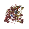


 PDBj
PDBj


 Assembly
 Sample preparation
 / Beamline: I04 / Wavelength: 0.9795 Å
 Processing