 Movie
Movie Controller


+ Open data

- Basic information
| Entry | Database: PDB / ID: 2f1o | ||||||
|---|---|---|---|---|---|---|---|
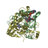
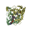
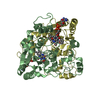
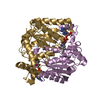
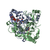
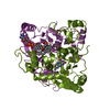
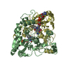
| Title | Crystal Structure of NQO1 with Dicoumarol | ||||||
 Components Components | NAD(P)H dehydrogenase [quinone] 1 | ||||||
 Keywords Keywords | OXIDOREDUCTASE/INHIBITOR / Protein inhibitor / Structural Genomics / Israel Structural Proteomics Center / ISPC / OXIDOREDUCTASE / OXIDOREDUCTASE-INHIBITOR complex | ||||||
| Function / homology |  Function and homology information Function and homology informationubiquinone metabolic process / vitamin E metabolic process / NAD(P)H dehydrogenase (quinone) / NADPH dehydrogenase (quinone) activity / cytochrome-b5 reductase activity, acting on NAD(P)H / vitamin K metabolic process / NADH dehydrogenase (quinone) (non-electrogenic) activity / NAD(P)H dehydrogenase (quinone) activity / NFE2L2 regulating anti-oxidant/detoxification enzymes / Regulation of ornithine decarboxylase (ODC) ...ubiquinone metabolic process / vitamin E metabolic process / NAD(P)H dehydrogenase (quinone) / NADPH dehydrogenase (quinone) activity / cytochrome-b5 reductase activity, acting on NAD(P)H / vitamin K metabolic process / NADH dehydrogenase (quinone) (non-electrogenic) activity / NAD(P)H dehydrogenase (quinone) activity / NFE2L2 regulating anti-oxidant/detoxification enzymes / Regulation of ornithine decarboxylase (ODC) / synaptic transmission, cholinergic / negative regulation of ferroptosis / nitric oxide biosynthetic process / removal of superoxide radicals / xenobiotic metabolic process / cell redox homeostasis / protein catabolic process / negative regulation of protein catabolic process / response to toxic substance / protein polyubiquitination / response to lipopolysaccharide / cellular response to oxidative stress / response to oxidative stress / innate immune response / synapse / RNA binding / identical protein binding / nucleus / cytosol / cytoplasm Similarity search - Function | ||||||
| Biological species |  Homo sapiens (human) Homo sapiens (human) | ||||||
| Method |  X-RAY DIFFRACTION / MOLECULAR REPLACEMENT / Resolution: 2.75 Å X-RAY DIFFRACTION / MOLECULAR REPLACEMENT / Resolution: 2.75 Å | ||||||
 Authors Authors | Shaul, Y. / Asher, G. / Dym, O. / Tsvetkov, P. / Adler, J. / Israel Structural Proteomics Center (ISPC) | ||||||
 Citation Citation | Journal: Biochemistry / Year: 2006 Title: The crystal structure of NAD(P)H quinone oxidoreductase 1 in complex with its potent inhibitor dicoumarol. Authors: Asher, G. / Dym, O. / Tsvetkov, P. / Adler, J. / Shaul, Y. | ||||||
| History |
|
- Structure visualization
| Structure viewer | Molecule: MolmilJmol/JSmol |
|---|
- Downloads & links
-Download
| PDBx/mmCIF format | 2f1o.cif.gz | 444.1 KB | Display | PDBx/mmCIF format |
|---|---|---|---|---|
| PDB format | pdb2f1o.ent.gz | 366.3 KB | Display | PDB format |
| PDBx/mmJSON format | 2f1o.json.gz | Tree view | PDBx/mmJSON format | |
| Others |  Other downloads Other downloads |
-Validation report
| Arichive directory | https://data.pdbj.org/pub/pdb/validation_reports/f1/2f1oftp://data.pdbj.org/pub/pdb/validation_reports/f1/2f1o | HTTPS FTP |
|---|
-Related structure data
| Related structure data |  1dxoS S: Starting model for refinement |
|---|---|
| Similar structure data | |
| Other databases |


-Links
 PDBj
PDBj
- Assembly
| Deposited unit | 
| ||||||||
|---|---|---|---|---|---|---|---|---|---|
| 1 | 
| ||||||||
| 2 | 
| ||||||||
| 3 | 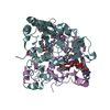
| ||||||||
| 4 | 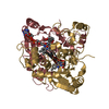
| ||||||||
| Unit cell |
| ||||||||
| Details | The biological assembly is a dimer which is in the asymmetric unit cell |
-Components
| #1: Protein | Mass: 30776.412 Da / Num. of mol.: 8 Source method: isolated from a genetically manipulated source Source: (gene. exp.) Homo sapiens (human) / Gene: NQO1, DIA4, NMOR1 / Production host:  References: UniProt: P15559, NAD(P)H dehydrogenase (quinone) #2: Chemical | ChemComp-DTC /   Mass: 336.295 Da / Num. of mol.: 8 / Source method: obtained synthetically / Formula: C19H12O6 Mass: 336.295 Da / Num. of mol.: 8 / Source method: obtained synthetically / Formula: C19H12O6#3: Chemical | ChemComp-FAD /   Mass: 785.550 Da / Num. of mol.: 8 / Source method: obtained synthetically / Formula: C27H33N9O15P2 / Comment: FAD*YM Mass: 785.550 Da / Num. of mol.: 8 / Source method: obtained synthetically / Formula: C27H33N9O15P2 / Comment: FAD*YM#4: Water | ChemComp-HOH / |  Mass: 18.015 Da / Num. of mol.: 122 / Source method: isolated from a natural source / Formula: H2O Mass: 18.015 Da / Num. of mol.: 122 / Source method: isolated from a natural source / Formula: H2O |
|---|
-Experimental details
-Experiment
| Experiment | Method: X-RAY DIFFRACTION / Number of used crystals: 1 |
|---|
- Sample preparation
| Crystal | Density Matthews: 2.55 Å3/Da / Density % sol: 51.71 % |
|---|---|
| Crystal grow | Temperature: 298 K / pH: 8.1 Details: 0.1M NaAcetate, 50mM NaTricine, 11% PEG 3350, 5mM Dicoumarol , pH 8.1, Microbatch, temperature 298K |
-Data collection
| Diffraction | Mean temperature: 100 K |
|---|---|
| Diffraction source | Source: ROTATING ANODE / Type: RIGAKU RU300 / Wavelength: 1.5418 |
| Radiation | Monochromator: YALE MIRRORS / Protocol: SINGLE WAVELENGTH / Monochromatic (M) / Laue (L): M / Scattering type: x-ray |
| Radiation wavelength | Wavelength: 1.5418 Å / Relative weight: 1 |
| Reflection | Resolution: 2.75→40 Å / Num. obs: 63333 / % possible obs: 96.9 % / Observed criterion σ(I): 2 |
| Reflection shell | Resolution: 2.75→2.8 Å / % possible all: 95.2 |
- Processing
| Software |
| ||||||||||||||||||||||||||||||||||||||||||||||||||||||||||||
|---|---|---|---|---|---|---|---|---|---|---|---|---|---|---|---|---|---|---|---|---|---|---|---|---|---|---|---|---|---|---|---|---|---|---|---|---|---|---|---|---|---|---|---|---|---|---|---|---|---|---|---|---|---|---|---|---|---|---|---|---|---|
| Refinement | Method to determine structure: MOLECULAR REPLACEMENT Starting model: 1DXO Resolution: 2.75→38.66 Å / σ(F): 0 / Stereochemistry target values: ENGH & HUBER
| ||||||||||||||||||||||||||||||||||||||||||||||||||||||||||||
| Refinement step | Cycle: LAST / Resolution: 2.75→38.66 Å
| ||||||||||||||||||||||||||||||||||||||||||||||||||||||||||||
| Refine LS restraints |
| ||||||||||||||||||||||||||||||||||||||||||||||||||||||||||||
| LS refinement shell | Resolution: 2.75→2.92 Å /
|
