 Movie
Movie Controller

[English] 日本語
 Yorodumi
- PDB-5a4k: Crystal structure of the R139W variant of human NAD(P)H:quinone o... -
+ Open data

- Basic information
| Entry | Database: PDB / ID: 5a4k | ||||||
|---|---|---|---|---|---|---|---|
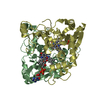
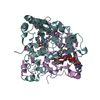
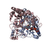
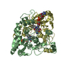
| Title | Crystal structure of the R139W variant of human NAD(P)H:quinone oxidoreductase | ||||||
 Components Components | NAD(P)H DEHYDROGENASE [QUINONE] 1 | ||||||
 Keywords Keywords | OXIDOREDUCTASE / NQO1 / FAD / FLAVOPROTEIN / OXIDATIVE STRESS / QUINONE REDUCTASE / DRUG METABOLISM / SINGLE AMINO ACID EXCHANGE | ||||||
| Function / homology |  Function and homology information Function and homology informationubiquinone metabolic process / vitamin E metabolic process / NAD(P)H dehydrogenase (quinone) / NADPH dehydrogenase (quinone) activity / cytochrome-b5 reductase activity, acting on NAD(P)H / vitamin K metabolic process / NADH dehydrogenase (quinone) (non-electrogenic) activity / NAD(P)H dehydrogenase (quinone) activity / NFE2L2 regulating anti-oxidant/detoxification enzymes / Regulation of ornithine decarboxylase (ODC) ...ubiquinone metabolic process / vitamin E metabolic process / NAD(P)H dehydrogenase (quinone) / NADPH dehydrogenase (quinone) activity / cytochrome-b5 reductase activity, acting on NAD(P)H / vitamin K metabolic process / NADH dehydrogenase (quinone) (non-electrogenic) activity / NAD(P)H dehydrogenase (quinone) activity / NFE2L2 regulating anti-oxidant/detoxification enzymes / Regulation of ornithine decarboxylase (ODC) / synaptic transmission, cholinergic / negative regulation of ferroptosis / nitric oxide biosynthetic process / removal of superoxide radicals / xenobiotic metabolic process / cell redox homeostasis / protein catabolic process / negative regulation of protein catabolic process / response to toxic substance / protein polyubiquitination / response to lipopolysaccharide / cellular response to oxidative stress / response to oxidative stress / innate immune response / synapse / RNA binding / identical protein binding / nucleus / cytosol / cytoplasm Similarity search - Function | ||||||
| Biological species |  HOMO SAPIENS (human) HOMO SAPIENS (human) | ||||||
| Method |  X-RAY DIFFRACTION / SYNCHROTRON / MOLECULAR REPLACEMENT / Resolution: 2.093 Å X-RAY DIFFRACTION / SYNCHROTRON / MOLECULAR REPLACEMENT / Resolution: 2.093 Å | ||||||
 Authors Authors | Lienhart, W.D. / Strandback, E. / Gudipati, V. / Uhl, M.K. / Rantase, D.M. / Zangger, K. / Gruber, K. / Macheroux, P. | ||||||
 Citation Citation | Journal: FEBS J. / Year: 2017 Title: Catalytic competence, structure and stability of the cancer-associated R139W variant of the human NAD(P)H:quinone oxidoreductase 1 (NQO1). Authors: Lienhart, W.D. / Strandback, E. / Gudipati, V. / Koch, K. / Binter, A. / Uhl, M.K. / Rantasa, D.M. / Bourgeois, B. / Madl, T. / Zangger, K. / Gruber, K. / Macheroux, P. | ||||||
| History |
|
- Structure visualization
| Structure viewer | Molecule: MolmilJmol/JSmol |
|---|
- Downloads & links
-Download
| PDBx/mmCIF format | 5a4k.cif.gz | 459.1 KB | Display | PDBx/mmCIF format |
|---|---|---|---|---|
| PDB format | pdb5a4k.ent.gz | 378.3 KB | Display | PDB format |
| PDBx/mmJSON format | 5a4k.json.gz | Tree view | PDBx/mmJSON format | |
| Others |  Other downloads Other downloads |
-Validation report
| Arichive directory | https://data.pdbj.org/pub/pdb/validation_reports/a4/5a4kftp://data.pdbj.org/pub/pdb/validation_reports/a4/5a4k | HTTPS FTP |
|---|
-Related structure data
| Related structure data |  1qbgS S: Starting model for refinement |
|---|---|
| Similar structure data |


-Links
 PDBj
PDBj
- Assembly
| Deposited unit | 
| ||||||||||||
|---|---|---|---|---|---|---|---|---|---|---|---|---|---|
| 1 | 
| ||||||||||||
| 2 | 
| ||||||||||||
| Unit cell |
| ||||||||||||
| Noncrystallographic symmetry (NCS) | NCS oper:
|
-Components
| #1: Protein | Mass: 33107.984 Da / Num. of mol.: 4 / Mutation: YES Source method: isolated from a genetically manipulated source Source: (gene. exp.) HOMO SAPIENS (human) / Plasmid: PET28A / Production host:  References: UniProt: P15559, NAD(P)H dehydrogenase (quinone) #2: Chemical | ChemComp-FAD /   Mass: 785.550 Da / Num. of mol.: 4 / Source method: obtained synthetically / Formula: C27H33N9O15P2 / Comment: FAD*YM Mass: 785.550 Da / Num. of mol.: 4 / Source method: obtained synthetically / Formula: C27H33N9O15P2 / Comment: FAD*YM#3: Chemical |   Mass: 209.240 Da / Num. of mol.: 2 / Source method: obtained synthetically / Formula: C8H19NO5 / Comment: pH buffer*YM Mass: 209.240 Da / Num. of mol.: 2 / Source method: obtained synthetically / Formula: C8H19NO5 / Comment: pH buffer*YM#4: Water | ChemComp-HOH / |  Mass: 18.015 Da / Num. of mol.: 1052 / Source method: isolated from a natural source / Formula: H2O Mass: 18.015 Da / Num. of mol.: 1052 / Source method: isolated from a natural source / Formula: H2OSequence details | N-TERMINAL HIS-TAG AND CLEAVAGE SITE R139W VARIANT | |
|---|
-Experimental details
-Experiment
| Experiment | Method: X-RAY DIFFRACTION / Number of used crystals: 1 |
|---|
- Sample preparation
| Crystal | Density Matthews: 2.3 Å3/Da / Density % sol: 46.57 % / Description: NONE |
|---|---|
| Crystal grow | Details: 200 MM LI2SO4, 100 MM BISTRIS (PH 6.5), 25% W/V PEG 3350, PROTEIN CONCENTRATION: 6.1 MG/ML |
-Data collection
| Diffraction | Mean temperature: 100 K |
|---|---|
| Diffraction source | Source: SYNCHROTRON / Site: SLS  / Beamline: X06DA / Wavelength: 1 / Beamline: X06DA / Wavelength: 1 |
| Detector | Type: DECTRIS PILATUS 2M-F / Detector: PIXEL / Details: TOROIDAL FOCUSING |
| Radiation | Monochromator: BARTELS MONOCHROMATOR, SI (111) / Protocol: SINGLE WAVELENGTH / Monochromatic (M) / Laue (L): M / Scattering type: x-ray |
| Radiation wavelength | Wavelength: 1 Å / Relative weight: 1 |
| Reflection | Resolution: 2.09→49.03 Å / Num. obs: 67587 / % possible obs: 97.1 % / Observed criterion σ(I): 0 / Redundancy: 2.9 % / Rmerge(I) obs: 0.08 / Net I/σ(I): 12.27 |
| Reflection shell | Resolution: 2.09→2.17 Å / Redundancy: 2.8 % / Rmerge(I) obs: 0.3 / Mean I/σ(I) obs: 4.1 / % possible all: 86.9 |
- Processing
| Software |
| |||||||||||||||||||||||||||||||||||||||||||||||||||||||||||||||||||||||||||||||||||||||||||||||||||||||||||||||||||||||||||||||||||||||||||||||||||||||||||||||||||||||||||||||||||||||||||||||||||||||||||||||||||||||||||||||||
|---|---|---|---|---|---|---|---|---|---|---|---|---|---|---|---|---|---|---|---|---|---|---|---|---|---|---|---|---|---|---|---|---|---|---|---|---|---|---|---|---|---|---|---|---|---|---|---|---|---|---|---|---|---|---|---|---|---|---|---|---|---|---|---|---|---|---|---|---|---|---|---|---|---|---|---|---|---|---|---|---|---|---|---|---|---|---|---|---|---|---|---|---|---|---|---|---|---|---|---|---|---|---|---|---|---|---|---|---|---|---|---|---|---|---|---|---|---|---|---|---|---|---|---|---|---|---|---|---|---|---|---|---|---|---|---|---|---|---|---|---|---|---|---|---|---|---|---|---|---|---|---|---|---|---|---|---|---|---|---|---|---|---|---|---|---|---|---|---|---|---|---|---|---|---|---|---|---|---|---|---|---|---|---|---|---|---|---|---|---|---|---|---|---|---|---|---|---|---|---|---|---|---|---|---|---|---|---|---|---|---|---|---|---|---|---|---|---|---|---|---|---|---|---|---|---|---|
| Refinement | Method to determine structure: MOLECULAR REPLACEMENT Starting model: PDB ENTRY 1QBG Resolution: 2.093→49.033 Å / SU ML: 0.22 / σ(F): 1.97 / Phase error: 20.69 / Stereochemistry target values: ML
| |||||||||||||||||||||||||||||||||||||||||||||||||||||||||||||||||||||||||||||||||||||||||||||||||||||||||||||||||||||||||||||||||||||||||||||||||||||||||||||||||||||||||||||||||||||||||||||||||||||||||||||||||||||||||||||||||
| Solvent computation | Shrinkage radii: 0.9 Å / VDW probe radii: 1.11 Å / Solvent model: FLAT BULK SOLVENT MODEL | |||||||||||||||||||||||||||||||||||||||||||||||||||||||||||||||||||||||||||||||||||||||||||||||||||||||||||||||||||||||||||||||||||||||||||||||||||||||||||||||||||||||||||||||||||||||||||||||||||||||||||||||||||||||||||||||||
| Refinement step | Cycle: LAST / Resolution: 2.093→49.033 Å
| |||||||||||||||||||||||||||||||||||||||||||||||||||||||||||||||||||||||||||||||||||||||||||||||||||||||||||||||||||||||||||||||||||||||||||||||||||||||||||||||||||||||||||||||||||||||||||||||||||||||||||||||||||||||||||||||||
| Refine LS restraints |
| |||||||||||||||||||||||||||||||||||||||||||||||||||||||||||||||||||||||||||||||||||||||||||||||||||||||||||||||||||||||||||||||||||||||||||||||||||||||||||||||||||||||||||||||||||||||||||||||||||||||||||||||||||||||||||||||||
| LS refinement shell |
| |||||||||||||||||||||||||||||||||||||||||||||||||||||||||||||||||||||||||||||||||||||||||||||||||||||||||||||||||||||||||||||||||||||||||||||||||||||||||||||||||||||||||||||||||||||||||||||||||||||||||||||||||||||||||||||||||
| Refinement TLS params. | Method: refined / Refine-ID: X-RAY DIFFRACTION
| |||||||||||||||||||||||||||||||||||||||||||||||||||||||||||||||||||||||||||||||||||||||||||||||||||||||||||||||||||||||||||||||||||||||||||||||||||||||||||||||||||||||||||||||||||||||||||||||||||||||||||||||||||||||||||||||||
| Refinement TLS group |
|
