 Movie
Movie Controller

[English] 日本語
 Yorodumi
- PDB-3jsx: X-ray Crystal structure of NAD(P)H: Quinone Oxidoreductase-1 (NQO... -
+ Open data

- Basic information
| Entry | Database: PDB / ID: 3jsx | ||||||
|---|---|---|---|---|---|---|---|
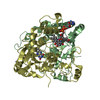
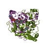
| Title | X-ray Crystal structure of NAD(P)H: Quinone Oxidoreductase-1 (NQO1) bound to the coumarin-based inhibitor AS1 | ||||||
 Components Components | NAD(P)H dehydrogenase [quinone] 1 | ||||||
 Keywords Keywords | OXIDOREDUCTASE / coumarin-based inhibitors / NQ01 / FAD / Flavoprotein / NAD / NADP | ||||||
| Function / homology |  Function and homology information Function and homology informationubiquinone metabolic process / vitamin E metabolic process / NAD(P)H dehydrogenase (quinone) / NADPH dehydrogenase (quinone) activity / cytochrome-b5 reductase activity, acting on NAD(P)H / vitamin K metabolic process / NADH dehydrogenase (quinone) (non-electrogenic) activity / NAD(P)H dehydrogenase (quinone) activity / response to caloric restriction / synaptic transmission, cholinergic ...ubiquinone metabolic process / vitamin E metabolic process / NAD(P)H dehydrogenase (quinone) / NADPH dehydrogenase (quinone) activity / cytochrome-b5 reductase activity, acting on NAD(P)H / vitamin K metabolic process / NADH dehydrogenase (quinone) (non-electrogenic) activity / NAD(P)H dehydrogenase (quinone) activity / response to caloric restriction / synaptic transmission, cholinergic / NFE2L2 regulating anti-oxidant/detoxification enzymes / Regulation of ornithine decarboxylase (ODC) / negative regulation of ferroptosis / nitric oxide biosynthetic process / xenobiotic metabolic process / removal of superoxide radicals / cell redox homeostasis / protein catabolic process / negative regulation of protein catabolic process / response to toxic substance / protein polyubiquitination / cellular response to oxidative stress / response to oxidative stress / response to lipopolysaccharide / innate immune response / synapse / RNA binding / identical protein binding / nucleus / cytoplasm / cytosol Similarity search - Function | ||||||
| Biological species |  Homo sapiens (human) Homo sapiens (human) | ||||||
| Method |  X-RAY DIFFRACTION / SYNCHROTRON / MOLECULAR REPLACEMENT / Resolution: 2.45 Å X-RAY DIFFRACTION / SYNCHROTRON / MOLECULAR REPLACEMENT / Resolution: 2.45 Å | ||||||
 Authors Authors | Dunstan, M.S. / Levy, C. / Leys, D. | ||||||
 Citation Citation | Journal: J.Med.Chem. / Year: 2009 Title: Synthesis and biological evaluation of coumarin-based inhibitors of NAD(P)H: quinone oxidoreductase-1 (NQO1). Authors: Nolan, K.A. / Doncaster, J.R. / Dunstan, M.S. / Scott, K.A. / Frenkel, A.D. / Siegel, D. / Ross, D. / Barnes, J. / Levy, C. / Leys, D. / Whitehead, R.C. / Stratford, I.J. / Bryce, R.A. | ||||||
| History |
|
- Structure visualization
| Structure viewer | Molecule: MolmilJmol/JSmol |
|---|
- Downloads & links
-Download
| PDBx/mmCIF format | 3jsx.cif.gz | 449.3 KB | Display | PDBx/mmCIF format |
|---|---|---|---|---|
| PDB format | pdb3jsx.ent.gz | 372 KB | Display | PDB format |
| PDBx/mmJSON format | 3jsx.json.gz | Tree view | PDBx/mmJSON format | |
| Others |  Other downloads Other downloads |
-Validation report
| Arichive directory | https://data.pdbj.org/pub/pdb/validation_reports/js/3jsxftp://data.pdbj.org/pub/pdb/validation_reports/js/3jsx | HTTPS FTP |
|---|
-Related structure data
| Related structure data |  2f1oS S: Starting model for refinement |
|---|---|
| Similar structure data |


-Links
 PDBj
PDBj
- Assembly
| Deposited unit | 
| ||||||||
|---|---|---|---|---|---|---|---|---|---|
| 1 | 
| ||||||||
| 2 | 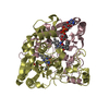
| ||||||||
| 3 | 
| ||||||||
| 4 | 
| ||||||||
| Unit cell |
|
-Components
| #1: Protein | Mass: 30776.412 Da / Num. of mol.: 8 Source method: isolated from a genetically manipulated source Source: (gene. exp.) Homo sapiens (human) / Gene: DIA4, NMOR1, NQO1, NQO1 cDNA / Plasmid: PKK 233-2 / Production host:  References: UniProt: P15559, NAD(P)H dehydrogenase (quinone) #2: Chemical | ChemComp-FAD /   Mass: 785.550 Da / Num. of mol.: 8 / Source method: obtained synthetically / Formula: C27H33N9O15P2 / Comment: FAD*YM Mass: 785.550 Da / Num. of mol.: 8 / Source method: obtained synthetically / Formula: C27H33N9O15P2 / Comment: FAD*YM#3: Chemical | ChemComp-CC2 /   Mass: 330.377 Da / Num. of mol.: 8 / Source method: obtained synthetically / Formula: C22H18O3 Mass: 330.377 Da / Num. of mol.: 8 / Source method: obtained synthetically / Formula: C22H18O3#4: Water | ChemComp-HOH / |  Mass: 18.015 Da / Num. of mol.: 434 / Source method: isolated from a natural source / Formula: H2O Mass: 18.015 Da / Num. of mol.: 434 / Source method: isolated from a natural source / Formula: H2O |
|---|
-Experimental details
-Experiment
| Experiment | Method: X-RAY DIFFRACTION / Number of used crystals: 1 |
|---|
- Sample preparation
| Crystal | Density Matthews: 2.87 Å3/Da / Density % sol: 57.08 % |
|---|---|
| Crystal grow | Temperature: 277 K / Method: vapor diffusion, sitting drop / pH: 8.5 Details: 2.4M ammonium sulfate, 0.1M Tris buffer pH 8.5, VAPOR DIFFUSION, SITTING DROP, temperature 277K |
-Data collection
| Diffraction | Mean temperature: 180 K |
|---|---|
| Diffraction source | Source: SYNCHROTRON / Site: Diamond  / Beamline: I02 / Wavelength: 0.9795 Å / Beamline: I02 / Wavelength: 0.9795 Å |
| Detector | Type: ADSC QUANTUM 315r / Detector: CCD / Date: May 9, 2009 |
| Radiation | Monochromator: Graphite / Protocol: SINGLE WAVELENGTH / Monochromatic (M) / Laue (L): M / Scattering type: x-ray |
| Radiation wavelength | Wavelength: 0.9795 Å / Relative weight: 1 |
| Reflection | Resolution: 2.45→47.954 Å / Num. all: 107399 / Num. obs: 100053 / % possible obs: 93.2 % / Observed criterion σ(F): 1.99 / Observed criterion σ(I): 1.99 / Redundancy: 4.1 % / Biso Wilson estimate: 53.46 Å2 / Rmerge(I) obs: 0.076 / Rsym value: 0.079 / Net I/σ(I): 13.98 |
| Reflection shell | Resolution: 2.45→2.51 Å / Redundancy: 2.7 % / Rmerge(I) obs: 0.458 / Mean I/σ(I) obs: 2.8 / Num. unique all: 6558 / % possible all: 84.4 |
- Processing
| Software |
| ||||||||||||||||||||||||||||||||||||||||||||||||||||||||||||||||||||||||||||||||||||||||||||||||||||||||||||||||||||||||||||||||||||||||||||||||||||||||||||||||||||||||||||||||||||||||||
|---|---|---|---|---|---|---|---|---|---|---|---|---|---|---|---|---|---|---|---|---|---|---|---|---|---|---|---|---|---|---|---|---|---|---|---|---|---|---|---|---|---|---|---|---|---|---|---|---|---|---|---|---|---|---|---|---|---|---|---|---|---|---|---|---|---|---|---|---|---|---|---|---|---|---|---|---|---|---|---|---|---|---|---|---|---|---|---|---|---|---|---|---|---|---|---|---|---|---|---|---|---|---|---|---|---|---|---|---|---|---|---|---|---|---|---|---|---|---|---|---|---|---|---|---|---|---|---|---|---|---|---|---|---|---|---|---|---|---|---|---|---|---|---|---|---|---|---|---|---|---|---|---|---|---|---|---|---|---|---|---|---|---|---|---|---|---|---|---|---|---|---|---|---|---|---|---|---|---|---|---|---|---|---|---|---|---|---|
| Refinement | Method to determine structure: MOLECULAR REPLACEMENT Starting model: 2F1O Resolution: 2.45→47.954 Å / SU ML: 0.15 / Isotropic thermal model: Isotropic / Cross valid method: THROUGHOUT / σ(F): 1.99 / σ(I): 2 / Phase error: 33.62 / Stereochemistry target values: ML
| ||||||||||||||||||||||||||||||||||||||||||||||||||||||||||||||||||||||||||||||||||||||||||||||||||||||||||||||||||||||||||||||||||||||||||||||||||||||||||||||||||||||||||||||||||||||||||
| Solvent computation | Shrinkage radii: 0.9 Å / VDW probe radii: 1.11 Å / Solvent model: FLAT BULK SOLVENT MODEL / Bsol: 53.469 Å2 / ksol: 0.334 e/Å3 | ||||||||||||||||||||||||||||||||||||||||||||||||||||||||||||||||||||||||||||||||||||||||||||||||||||||||||||||||||||||||||||||||||||||||||||||||||||||||||||||||||||||||||||||||||||||||||
| Displacement parameters | Biso mean: 53.6 Å2
| ||||||||||||||||||||||||||||||||||||||||||||||||||||||||||||||||||||||||||||||||||||||||||||||||||||||||||||||||||||||||||||||||||||||||||||||||||||||||||||||||||||||||||||||||||||||||||
| Refine analyze | Luzzati coordinate error obs: 0.15 Å | ||||||||||||||||||||||||||||||||||||||||||||||||||||||||||||||||||||||||||||||||||||||||||||||||||||||||||||||||||||||||||||||||||||||||||||||||||||||||||||||||||||||||||||||||||||||||||
| Refinement step | Cycle: LAST / Resolution: 2.45→47.954 Å
| ||||||||||||||||||||||||||||||||||||||||||||||||||||||||||||||||||||||||||||||||||||||||||||||||||||||||||||||||||||||||||||||||||||||||||||||||||||||||||||||||||||||||||||||||||||||||||
| Refine LS restraints |
| ||||||||||||||||||||||||||||||||||||||||||||||||||||||||||||||||||||||||||||||||||||||||||||||||||||||||||||||||||||||||||||||||||||||||||||||||||||||||||||||||||||||||||||||||||||||||||
| LS refinement shell | Refine-ID: X-RAY DIFFRACTION / Total num. of bins used: 30
|
