 Movie
Movie Controller

[English] 日本語
 Yorodumi
- PDB-5mkv: Crystal Structure of Human Dihydropyrimidinease-like 2 (DPYSL2A)/... -
+ Open data

- Basic information
| Entry | Database: PDB / ID: 5mkv | ||||||
|---|---|---|---|---|---|---|---|
| Title | Crystal Structure of Human Dihydropyrimidinease-like 2 (DPYSL2A)/Collapsin Response Mediator Protein (CRMP2) residues 13-516 | ||||||
 Components Components | Dihydropyrimidinase-related protein 2 | ||||||
 Keywords Keywords | HYDROLASE / CRMP2 / Ovarian cancer / Microtubule associated protein | ||||||
| Function / homology |  Function and homology information Function and homology informationdihydropyrimidinase activity / hydrolase activity, acting on carbon-nitrogen (but not peptide) bonds, in cyclic amides / CRMPs in Sema3A signaling / nucleobase-containing compound metabolic process / Recycling pathway of L1 / cytoskeleton organization / endocytosis / mitotic spindle / nervous system development / microtubule cytoskeleton ...dihydropyrimidinase activity / hydrolase activity, acting on carbon-nitrogen (but not peptide) bonds, in cyclic amides / CRMPs in Sema3A signaling / nucleobase-containing compound metabolic process / Recycling pathway of L1 / cytoskeleton organization / endocytosis / mitotic spindle / nervous system development / microtubule cytoskeleton / cell differentiation / cilium / signal transduction / extracellular exosome / identical protein binding / plasma membrane / cytosol Similarity search - Function | ||||||
| Biological species |  Homo sapiens (human) Homo sapiens (human) | ||||||
| Method |  X-RAY DIFFRACTION / SYNCHROTRON / MOLECULAR REPLACEMENT / Resolution: 1.8 Å X-RAY DIFFRACTION / SYNCHROTRON / MOLECULAR REPLACEMENT / Resolution: 1.8 Å | ||||||
 Authors Authors | Sethi, R. / Zheng, Y. / Krojer, T. / Velupillai, S. / Arrowsmith, C.H. / Edwards, A.M. / Bountra, C. / Ahmed, A.A. / von Delft, F. | ||||||
 Citation Citation | Journal: Nat Commun / Year: 2018 Title: Tuning microtubule dynamics to enhance cancer therapy by modulating FER-mediated CRMP2 phosphorylation. Authors: Zheng, Y. / Sethi, R. / Mangala, L.S. / Taylor, C. / Goldsmith, J. / Wang, M. / Masuda, K. / Karaminejadranjbar, M. / Mannion, D. / Miranda, F. / Herrero-Gonzalez, S. / Hellner, K. / Chen, F. ...Authors: Zheng, Y. / Sethi, R. / Mangala, L.S. / Taylor, C. / Goldsmith, J. / Wang, M. / Masuda, K. / Karaminejadranjbar, M. / Mannion, D. / Miranda, F. / Herrero-Gonzalez, S. / Hellner, K. / Chen, F. / Alsaadi, A. / Albukhari, A. / Fotso, D.C. / Yau, C. / Jiang, D. / Pradeep, S. / Rodriguez-Aguayo, C. / Lopez-Berestein, G. / Knapp, S. / Gray, N.S. / Campo, L. / Myers, K.A. / Dhar, S. / Ferguson, D. / Bast, R.C. / Sood, A.K. / von Delft, F. / Ahmed, A.A. | ||||||
| History |
|
- Structure visualization
| Structure viewer | Molecule: MolmilJmol/JSmol |
|---|
- Downloads & links
-Download
| PDBx/mmCIF format | 5mkv.cif.gz | 389 KB | Display | PDBx/mmCIF format |
|---|---|---|---|---|
| PDB format | pdb5mkv.ent.gz | 315.1 KB | Display | PDB format |
| PDBx/mmJSON format | 5mkv.json.gz | Tree view | PDBx/mmJSON format | |
| Others |  Other downloads Other downloads |
-Validation report
| Arichive directory | https://data.pdbj.org/pub/pdb/validation_reports/mk/5mkvftp://data.pdbj.org/pub/pdb/validation_reports/mk/5mkv | HTTPS FTP |
|---|
-Related structure data
| Related structure data |  5mleC 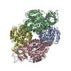 2gseS S: Starting model for refinement C: citing same article ( |
|---|---|
| Similar structure data |


-Links
 PDBj
PDBj


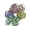
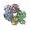
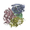
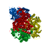
- Assembly
| Deposited unit | 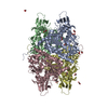
| ||||||||
|---|---|---|---|---|---|---|---|---|---|
| 1 |
| ||||||||
| Unit cell |
|
-Components
| #1: Protein | Mass: 55104.156 Da / Num. of mol.: 4 Source method: isolated from a genetically manipulated source Source: (gene. exp.) Homo sapiens (human) / Gene: DPYSL2, CRMP2, ULIP2 / Production host:  #2: Chemical | ChemComp-EDO /   Mass: 62.068 Da / Num. of mol.: 22 / Source method: obtained synthetically / Formula: C2H6O2 Mass: 62.068 Da / Num. of mol.: 22 / Source method: obtained synthetically / Formula: C2H6O2#3: Water | ChemComp-HOH / |  Mass: 18.015 Da / Num. of mol.: 596 / Source method: isolated from a natural source / Formula: H2O Mass: 18.015 Da / Num. of mol.: 596 / Source method: isolated from a natural source / Formula: H2O |
|---|
-Experimental details
-Experiment
| Experiment | Method: X-RAY DIFFRACTION / Number of used crystals: 1 |
|---|
- Sample preparation
| Crystal | Density Matthews: 2.49 Å3/Da / Density % sol: 50.62 % |
|---|---|
| Crystal grow | Temperature: 293 K / Method: vapor diffusion, sitting drop / pH: 5.5 Details: 0.2M ammonium acetate, 30% PEG 4000 and 0.1M citrate buffer pH 5.5 |
-Data collection
| Diffraction | Mean temperature: 100 K |
|---|---|
| Diffraction source | Source: SYNCHROTRON / Site: Diamond  / Beamline: I04-1 / Wavelength: 0.92 Å / Beamline: I04-1 / Wavelength: 0.92 Å |
| Detector | Type: DECTRIS PILATUS 6M-F / Detector: PIXEL / Date: Feb 4, 2015 |
| Radiation | Protocol: SINGLE WAVELENGTH / Monochromatic (M) / Laue (L): M / Scattering type: x-ray |
| Radiation wavelength | Wavelength: 0.92 Å / Relative weight: 1 |
| Reflection | Resolution: 1.8→19.87 Å / Num. obs: 194435 / % possible obs: 97.71 % / Redundancy: 3.6 % / Net I/σ(I): 15.84 |
- Processing
| Software |
| ||||||||||||||||||||||||||||||||||||||||||||||||||||||||||||||||||||||||||||||||||||||||||||||||||||||||||||||||||||||||||||||||||||||||||||||||||||||||||||||||||||||||||||||||||||||
|---|---|---|---|---|---|---|---|---|---|---|---|---|---|---|---|---|---|---|---|---|---|---|---|---|---|---|---|---|---|---|---|---|---|---|---|---|---|---|---|---|---|---|---|---|---|---|---|---|---|---|---|---|---|---|---|---|---|---|---|---|---|---|---|---|---|---|---|---|---|---|---|---|---|---|---|---|---|---|---|---|---|---|---|---|---|---|---|---|---|---|---|---|---|---|---|---|---|---|---|---|---|---|---|---|---|---|---|---|---|---|---|---|---|---|---|---|---|---|---|---|---|---|---|---|---|---|---|---|---|---|---|---|---|---|---|---|---|---|---|---|---|---|---|---|---|---|---|---|---|---|---|---|---|---|---|---|---|---|---|---|---|---|---|---|---|---|---|---|---|---|---|---|---|---|---|---|---|---|---|---|---|---|---|
| Refinement | Method to determine structure: MOLECULAR REPLACEMENT Starting model: 2GSE Resolution: 1.8→19.87 Å / Cor.coef. Fo:Fc: 0.965 / Cor.coef. Fo:Fc free: 0.951 / SU B: 2.434 / SU ML: 0.075 / Cross valid method: THROUGHOUT / ESU R: 0.11 / ESU R Free: 0.107 / Details: HYDROGENS HAVE BEEN ADDED IN THE RIDING POSITIONS
| ||||||||||||||||||||||||||||||||||||||||||||||||||||||||||||||||||||||||||||||||||||||||||||||||||||||||||||||||||||||||||||||||||||||||||||||||||||||||||||||||||||||||||||||||||||||
| Solvent computation | Ion probe radii: 0.8 Å / Shrinkage radii: 0.8 Å / VDW probe radii: 1.2 Å | ||||||||||||||||||||||||||||||||||||||||||||||||||||||||||||||||||||||||||||||||||||||||||||||||||||||||||||||||||||||||||||||||||||||||||||||||||||||||||||||||||||||||||||||||||||||
| Displacement parameters | Biso mean: 25.454 Å2
| ||||||||||||||||||||||||||||||||||||||||||||||||||||||||||||||||||||||||||||||||||||||||||||||||||||||||||||||||||||||||||||||||||||||||||||||||||||||||||||||||||||||||||||||||||||||
| Refinement step | Cycle: 1 / Resolution: 1.8→19.87 Å
| ||||||||||||||||||||||||||||||||||||||||||||||||||||||||||||||||||||||||||||||||||||||||||||||||||||||||||||||||||||||||||||||||||||||||||||||||||||||||||||||||||||||||||||||||||||||
| Refine LS restraints |
|