[English] 日本語
Yorodumi
- PDB-2vm8: Human CRMP-2 crystallised in the presence of Mg -

+
Open data


ID or keywords:

Loading...

-
Basic information

Entry
Database: PDB / ID: 2vm8
TitleHuman CRMP-2 crystallised in the presence of Mg
ComponentsDIHYDROPYRIMIDINASE-RELATED PROTEIN 2
KeywordsSIGNALING PROTEIN / NEUROGENESIS / PHOSPHOPROTEIN / DIFFERENTIATION / CRMP / CYTOPLASM / TIM BARREL / POLYMORPHISM / AXONAL PATHFINDING / DEVELOPMENTAL PROTEIN
Function / homology
Function and homology information


dihydropyrimidinase activity / hydrolase activity, acting on carbon-nitrogen (but not peptide) bonds, in cyclic amides / CRMPs in Sema3A signaling / nucleobase-containing compound metabolic process / Recycling pathway of L1 / cytoskeleton organization / endocytosis / nervous system development / cell differentiation / cytoskeleton ...dihydropyrimidinase activity / hydrolase activity, acting on carbon-nitrogen (but not peptide) bonds, in cyclic amides / CRMPs in Sema3A signaling / nucleobase-containing compound metabolic process / Recycling pathway of L1 / cytoskeleton organization / endocytosis / nervous system development / cell differentiation / cytoskeleton / signal transduction / extracellular exosome / identical protein binding / plasma membrane / cytosol
Similarity search - Function
Hydantoinase/dihydropyrimidinase / Urease, subunit C; domain 1 / Urease, subunit C, domain 1 / Amidohydrolase family / Metal-dependent hydrolase, composite domain superfamily / Amidohydrolase-related / Metal-dependent hydrolases / Metal-dependent hydrolase / Roll / TIM Barrel ...Hydantoinase/dihydropyrimidinase / Urease, subunit C; domain 1 / Urease, subunit C, domain 1 / Amidohydrolase family / Metal-dependent hydrolase, composite domain superfamily / Amidohydrolase-related / Metal-dependent hydrolases / Metal-dependent hydrolase / Roll / TIM Barrel / Alpha-Beta Barrel / Mainly Beta / Alpha Beta
Similarity search - Domain/homology
Dihydropyrimidinase-related protein 2
Similarity search - Component
Biological speciesHOMO SAPIENS (human)
MethodX-RAY DIFFRACTION / SYNCHROTRON / MOLECULAR REPLACEMENT / Resolution: 1.9 Å
AuthorsKursula, P.
CitationJournal: FEBS J. / Year: 2008
Title: Crystal and Solution Structure, Stability and Post- Translational Modifications of Collapsin Response Mediator Protein 2.
Authors: Majava, V. / Loytynoja, N. / Chen, W.Q. / Lubec, G. / Kursula, P.
History
DepositionJan 24, 2008Deposition site: PDBE / Processing site: PDBE
Revision 1.0Aug 26, 2008Provider: repository / Type: Initial release
Revision 1.1May 8, 2011Group: Version format compliance
Revision 1.2Jul 13, 2011Group: Version format compliance
Revision 1.3Jan 17, 2018Group: Data collection / Category: diffrn_source / Item: _diffrn_source.pdbx_synchrotron_site
Revision 1.4Dec 13, 2023Group: Data collection / Database references ...Data collection / Database references / Derived calculations / Other / Refinement description
Category: chem_comp_atom / chem_comp_bond ...chem_comp_atom / chem_comp_bond / database_2 / pdbx_database_status / pdbx_initial_refinement_model / pdbx_struct_conn_angle / struct_conn / struct_site
Item: _database_2.pdbx_DOI / _database_2.pdbx_database_accession ..._database_2.pdbx_DOI / _database_2.pdbx_database_accession / _pdbx_database_status.status_code_sf / _pdbx_struct_conn_angle.ptnr1_auth_seq_id / _pdbx_struct_conn_angle.ptnr3_auth_seq_id / _pdbx_struct_conn_angle.value / _struct_conn.pdbx_dist_value / _struct_conn.ptnr2_auth_seq_id / _struct_site.pdbx_auth_asym_id / _struct_site.pdbx_auth_comp_id / _struct_site.pdbx_auth_seq_id
Remark 700 SHEET THE SHEET STRUCTURE OF THIS MOLECULE IS BIFURCATED. IN ORDER TO REPRESENT THIS FEATURE IN ... SHEET THE SHEET STRUCTURE OF THIS MOLECULE IS BIFURCATED. IN ORDER TO REPRESENT THIS FEATURE IN THE SHEET RECORDS BELOW, TWO SHEETS ARE DEFINED.

-
Structure visualization

Structure viewerMolecule:
MolmilJmol/JSmol

Downloads & links

-
Assembly

Deposited unit
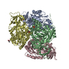
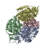
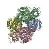
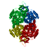
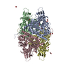
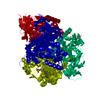
A: DIHYDROPYRIMIDINASE-RELATED PROTEIN 2
B: DIHYDROPYRIMIDINASE-RELATED PROTEIN 2
C: DIHYDROPYRIMIDINASE-RELATED PROTEIN 2
D: DIHYDROPYRIMIDINASE-RELATED PROTEIN 2
hetero molecules


Theoretical massNumber of molelcules
Total (without water)220,4617
Polymers220,3884
Non-polymers733
Water32,3011793
1


  • Idetical with deposited unit
  • defined by software
TypeNameSymmetry operationNumber
identity operation1_555x,y,z1
Buried area10540 Å2
ΔGint-34 kcal/mol
Surface area77000 Å2
MethodPQS
Unit cell
Length a, b, c (Å)86.510, 126.240, 209.750
Angle α, β, γ (deg.)90.00, 90.00, 90.00
Int Tables number19
Space group name H-MP212121

-
Components

#1: Protein
DIHYDROPYRIMIDINASE-RELATED PROTEIN 2 / COLLAPSIN RESPONSE MEDIATOR PROTEIN 2 / DRP-2 / CRMP-2 / N2A3


Mass: 55096.969 Da / Num. of mol.: 4 / Fragment: RESIDUES 13-490
Source method: isolated from a genetically manipulated source
Source: (gene. exp.) HOMO SAPIENS (human) / Plasmid: PNIC-BSA4 / Production host: ESCHERICHIA COLI (E. coli) / Strain (production host): BL21(DE3) / References: UniProt: Q16555
#2: Chemical ChemComp-MG / MAGNESIUM ION


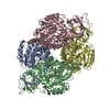
Mass: 24.305 Da / Num. of mol.: 3 / Source method: obtained synthetically / Formula: Mg
#3: Water ChemComp-HOH / water / Water


Mass: 18.015 Da / Num. of mol.: 1793 / Source method: isolated from a natural source / Formula: H2O

-
Experimental details

-
Experiment

ExperimentMethod: X-RAY DIFFRACTION / Number of used crystals: 1

-
Sample preparation

CrystalDensity Matthews: 2.47 Å3/Da / Density % sol: 49.88 % / Description: NONE

-
Data collection

DiffractionMean temperature: 100 K
Diffraction sourceSource: SYNCHROTRON / Site: MAX II / Beamline: I911-2 / Wavelength: 1.0408
DetectorType: MARRESEARCH / Detector: CCD / Date: Oct 6, 2006
RadiationProtocol: SINGLE WAVELENGTH / Monochromatic (M) / Laue (L): M / Scattering type: x-ray
Radiation wavelengthWavelength: 1.0408 Å / Relative weight: 1
ReflectionResolution: 1.9→20 Å / Num. obs: 170498 / % possible obs: 94.2 % / Observed criterion σ(I): -3 / Redundancy: 6 % / Biso Wilson estimate: 22 Å2 / Rmerge(I) obs: 0.12 / Net I/σ(I): 11.4
Reflection shellResolution: 1.9→2 Å / Redundancy: 5.5 % / Rmerge(I) obs: 0.47 / Mean I/σ(I) obs: 3.8 / % possible all: 83.7

-
Processing

Software
NameVersionClassification
REFMAC5.3.0028refinement
XDSdata reduction
XSCALEdata scaling
MOLREPphasing
RefinementMethod to determine structure: MOLECULAR REPLACEMENT
Starting model: PDB ENTRY 2GSE
Resolution: 1.9→20 Å / Cor.coef. Fo:Fc: 0.915 / Cor.coef. Fo:Fc free: 0.847 / SU B: 7.571 / SU ML: 0.211 / Cross valid method: THROUGHOUT / ESU R: 0.209 / ESU R Free: 0.204 / Stereochemistry target values: MAXIMUM LIKELIHOOD
Details: HYDROGENS HAVE BEEN ADDED IN THE RIDING POSITIONS. DUE TO PSEUDOTRANSLATIONAL SYMMETRY, THE R FACTORS REMAINED HIGHER THAN EXPECTED DURING REFINEMENT.
RfactorNum. reflection% reflectionSelection details
Rfree0.325 8539 5 %RANDOM
Rwork0.256 ---
obs0.259 162237 94.5 %-
Solvent computationIon probe radii: 0.8 Å / Shrinkage radii: 0.8 Å / VDW probe radii: 1.4 Å / Solvent model: BABINET MODEL WITH MASK
Displacement parametersBiso mean: 17.39 Å2
Baniso -1Baniso -2Baniso -3
1--2.04 Å20 Å20 Å2
2---0.41 Å20 Å2
3---2.45 Å2
Refinement stepCycle: LAST / Resolution: 1.9→20 Å
ProteinNucleic acidLigandSolventTotal
Num. atoms14674 0 3 1793 16470
Refine LS restraints
Refine-IDTypeDev idealDev ideal targetNumber
X-RAY DIFFRACTIONr_bond_refined_d0.0160.02215057
X-RAY DIFFRACTIONr_bond_other_d0.0010.0210053
X-RAY DIFFRACTIONr_angle_refined_deg1.5791.95120454
X-RAY DIFFRACTIONr_angle_other_deg0.984324649
X-RAY DIFFRACTIONr_dihedral_angle_1_deg6.64551929
X-RAY DIFFRACTIONr_dihedral_angle_2_deg38.43524.797665
X-RAY DIFFRACTIONr_dihedral_angle_3_deg16.061152559
X-RAY DIFFRACTIONr_dihedral_angle_4_deg17.6451577
X-RAY DIFFRACTIONr_chiral_restr0.0870.22311
X-RAY DIFFRACTIONr_gen_planes_refined0.0050.0216902
X-RAY DIFFRACTIONr_gen_planes_other0.0010.022907
X-RAY DIFFRACTIONr_nbd_refined0.2260.23900
X-RAY DIFFRACTIONr_nbd_other0.2090.211546
X-RAY DIFFRACTIONr_nbtor_refined0.1840.27390
X-RAY DIFFRACTIONr_nbtor_other0.090.28123
X-RAY DIFFRACTIONr_xyhbond_nbd_refined0.2450.21472
X-RAY DIFFRACTIONr_xyhbond_nbd_other
X-RAY DIFFRACTIONr_metal_ion_refined
X-RAY DIFFRACTIONr_metal_ion_other
X-RAY DIFFRACTIONr_symmetry_vdw_refined0.1830.27
X-RAY DIFFRACTIONr_symmetry_vdw_other0.2360.238
X-RAY DIFFRACTIONr_symmetry_hbond_refined0.1760.213
X-RAY DIFFRACTIONr_symmetry_hbond_other
X-RAY DIFFRACTIONr_symmetry_metal_ion_refined
X-RAY DIFFRACTIONr_symmetry_metal_ion_other
X-RAY DIFFRACTIONr_mcbond_it1.471212242
X-RAY DIFFRACTIONr_mcbond_other
X-RAY DIFFRACTIONr_mcangle_it1.891315384
X-RAY DIFFRACTIONr_mcangle_other
X-RAY DIFFRACTIONr_scbond_it2.6746371
X-RAY DIFFRACTIONr_scbond_other
X-RAY DIFFRACTIONr_scangle_it3.42855056
X-RAY DIFFRACTIONr_scangle_other
X-RAY DIFFRACTIONr_long_range_B_refined
X-RAY DIFFRACTIONr_long_range_B_other
X-RAY DIFFRACTIONr_rigid_bond_restr
X-RAY DIFFRACTIONr_sphericity_free
X-RAY DIFFRACTIONr_sphericity_bonded
LS refinement shellResolution: 1.9→1.95 Å / Total num. of bins used: 20 /
RfactorNum. reflection
Rfree0.419 535
Rwork0.384 10173

+
About Yorodumi

-
News

-
Feb 9, 2022. New format data for meta-information of EMDB entries

New format data for meta-information of EMDB entries

  • Version 3 of the EMDB header file is now the official format.
  • The previous official version 1.9 will be removed from the archive.

Related info.:EMDB header

External links:wwPDB to switch to version 3 of the EMDB data model

-
Aug 12, 2020. Covid-19 info

Covid-19 info

URL: https://pdbj.org/emnavi/covid19.php

New page: Covid-19 featured information page in EM Navigator.

Related info.:Covid-19 info / Mar 5, 2020. Novel coronavirus structure data

+
Mar 5, 2020. Novel coronavirus structure data

Novel coronavirus structure data

Related info.:Yorodumi Speices / Aug 12, 2020. Covid-19 info

External links:COVID-19 featured content - PDBj / Molecule of the Month (242):Coronavirus Proteases

+
Jan 31, 2019. EMDB accession codes are about to change! (news from PDBe EMDB page)

EMDB accession codes are about to change! (news from PDBe EMDB page)

  • The allocation of 4 digits for EMDB accession codes will soon come to an end. Whilst these codes will remain in use, new EMDB accession codes will include an additional digit and will expand incrementally as the available range of codes is exhausted. The current 4-digit format prefixed with “EMD-” (i.e. EMD-XXXX) will advance to a 5-digit format (i.e. EMD-XXXXX), and so on. It is currently estimated that the 4-digit codes will be depleted around Spring 2019, at which point the 5-digit format will come into force.
  • The EM Navigator/Yorodumi systems omit the EMD- prefix.

Related info.:Q: What is EMD? / ID/Accession-code notation in Yorodumi/EM Navigator

External links:EMDB Accession Codes are Changing Soon! / Contact to PDBj

+
Jul 12, 2017. Major update of PDB

Major update of PDB

  • wwPDB released updated PDB data conforming to the new PDBx/mmCIF dictionary.
  • This is a major update changing the version number from 4 to 5, and with Remediation, in which all the entries are updated.
  • In this update, many items about electron microscopy experimental information are reorganized (e.g. em_software).
  • Now, EM Navigator and Yorodumi are based on the updated data.

External links:wwPDB Remediation / Enriched Model Files Conforming to OneDep Data Standards Now Available in the PDB FTP Archive

-
Yorodumi

Thousand views of thousand structures

  • Yorodumi is a browser for structure data from EMDB, PDB, SASBDB, etc.
  • This page is also the successor to EM Navigator detail page, and also detail information page/front-end page for Omokage search.
  • The word "yorodu" (or yorozu) is an old Japanese word meaning "ten thousand". "mi" (miru) is to see.

Related info.:EMDB / PDB / SASBDB / Comparison of 3 databanks / Yorodumi Search / Aug 31, 2016. New EM Navigator & Yorodumi / Yorodumi Papers / Jmol/JSmol / Function and homology information / Changes in new EM Navigator and Yorodumi

Read more