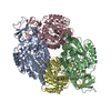
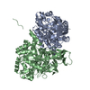
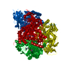
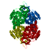
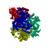
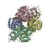
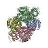
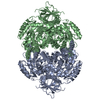
[English] 日本語
Yorodumi
- PDB-4cnu: CRYSTAL STRUCTURE OF WT HUMAN CRMP-4 from lattice translocation -

+
Open data


ID or keywords:

Loading...

-
Basic information

Entry
Database: PDB / ID: 4cnu
TitleCRYSTAL STRUCTURE OF WT HUMAN CRMP-4 from lattice translocation
ComponentsDIHYDROPYRIMIDINASE-LIKE 3
KeywordsSIGNALING PROTEIN / NEUROGENESIS / AXONAL OUTGROWTH / DEVELOPMENTAL PROTEIN
Function / homology
Function and homology information


hydrolase activity, acting on carbon-nitrogen (but not peptide) bonds, in cyclic amides / chondroitin sulfate binding / filamin binding / actin crosslink formation / exocytic vesicle / CRMPs in Sema3A signaling / positive regulation of filopodium assembly / cellular response to cytokine stimulus / filamentous actin / actin filament bundle assembly ...hydrolase activity, acting on carbon-nitrogen (but not peptide) bonds, in cyclic amides / chondroitin sulfate binding / filamin binding / actin crosslink formation / exocytic vesicle / CRMPs in Sema3A signaling / positive regulation of filopodium assembly / cellular response to cytokine stimulus / filamentous actin / actin filament bundle assembly / neuron development / response to axon injury / negative regulation of cell migration / positive regulation of neuron projection development / SH3 domain binding / negative regulation of neuron projection development / lamellipodium / cell body / growth cone / synapse / extracellular space / identical protein binding / cytosol
Similarity search - Function
Hydantoinase/dihydropyrimidinase / Urease, subunit C; domain 1 / Urease, subunit C, domain 1 / Amidohydrolase family / Metal-dependent hydrolase, composite domain superfamily / Amidohydrolase-related / Metal-dependent hydrolases / Metal-dependent hydrolase / Roll / TIM Barrel ...Hydantoinase/dihydropyrimidinase / Urease, subunit C; domain 1 / Urease, subunit C, domain 1 / Amidohydrolase family / Metal-dependent hydrolase, composite domain superfamily / Amidohydrolase-related / Metal-dependent hydrolases / Metal-dependent hydrolase / Roll / TIM Barrel / Alpha-Beta Barrel / Mainly Beta / Alpha Beta
Similarity search - Domain/homology
Dihydropyrimidinase-related protein 3
Similarity search - Component
Biological speciesHOMO SAPIENS (human)
MethodX-RAY DIFFRACTION / SYNCHROTRON / MOLECULAR REPLACEMENT / Resolution: 2.8 Å
AuthorsPonnusamy, R. / Lebedev, A. / Lohkamp, B.
CitationJournal: Acta Crystallogr.,Sect.D / Year: 2014
Title: Crystal Structure of Human Crmp-4: Correction of Intensities for Lattice-Translocation Disorder
Authors: Ponnusamy, R. / Lebedev, A. / Pahlow, S. / Lohkamp, B.
History
DepositionJan 24, 2014Deposition site: PDBE / Processing site: PDBE
Revision 1.0Jun 18, 2014Provider: repository / Type: Initial release
Revision 1.1Jan 17, 2018Group: Data collection / Category: diffrn_source / Item: _diffrn_source.pdbx_synchrotron_site
Revision 1.2Dec 20, 2023Group: Data collection / Database references ...Data collection / Database references / Other / Refinement description
Category: chem_comp_atom / chem_comp_bond ...chem_comp_atom / chem_comp_bond / database_2 / pdbx_database_status / pdbx_initial_refinement_model / struct_ncs_dom_lim
Item: _database_2.pdbx_DOI / _database_2.pdbx_database_accession ..._database_2.pdbx_DOI / _database_2.pdbx_database_accession / _pdbx_database_status.status_code_sf / _struct_ncs_dom_lim.beg_auth_comp_id / _struct_ncs_dom_lim.beg_label_asym_id / _struct_ncs_dom_lim.beg_label_comp_id / _struct_ncs_dom_lim.beg_label_seq_id / _struct_ncs_dom_lim.end_auth_comp_id / _struct_ncs_dom_lim.end_label_asym_id / _struct_ncs_dom_lim.end_label_comp_id / _struct_ncs_dom_lim.end_label_seq_id

-
Structure visualization

Structure viewerMolecule:
MolmilJmol/JSmol

Downloads & links

-
Assembly

Deposited unit
A: DIHYDROPYRIMIDINASE-LIKE 3
B: DIHYDROPYRIMIDINASE-LIKE 3


Theoretical massNumber of molelcules
Total (without water)124,0982
Polymers124,0982
Non-polymers00
Water0
1
A: DIHYDROPYRIMIDINASE-LIKE 3

A: DIHYDROPYRIMIDINASE-LIKE 3

B: DIHYDROPYRIMIDINASE-LIKE 3

B: DIHYDROPYRIMIDINASE-LIKE 3


Theoretical massNumber of molelcules
Total (without water)248,1974
Polymers248,1974
Non-polymers00
Water0
TypeNameSymmetry operationNumber
identity operation1_555x,y,z1
crystal symmetry operation2_555-x,-y,z1
crystal symmetry operation3_546-x+1/2,y-1/2,-z+11
crystal symmetry operation4_456x-1/2,-y+1/2,-z+11
Buried area12900 Å2
ΔGint-52.3 kcal/mol
Surface area62230 Å2
MethodPISA
2
B: DIHYDROPYRIMIDINASE-LIKE 3

B: DIHYDROPYRIMIDINASE-LIKE 3

A: DIHYDROPYRIMIDINASE-LIKE 3

A: DIHYDROPYRIMIDINASE-LIKE 3


Theoretical massNumber of molelcules
Total (without water)248,1974
Polymers248,1974
Non-polymers00
Water0
TypeNameSymmetry operationNumber
identity operation1_555x,y,z1
crystal symmetry operation2_665-x+1,-y+1,z1
crystal symmetry operation4_556x+1/2,-y+1/2,-z+11
crystal symmetry operation3_556-x+1/2,y+1/2,-z+11
Buried area12900 Å2
ΔGint-52.3 kcal/mol
Surface area62230 Å2
MethodPISA
Unit cell
Length a, b, c (Å)96.240, 108.220, 118.190
Angle α, β, γ (deg.)90.00, 90.00, 90.00
Int Tables number18
Space group name H-MP21212
Noncrystallographic symmetry (NCS)NCS domain:
IDEns-IDDetails
11A
21B

NCS domain segments:

Component-ID: 0 / Ens-ID: 1 / Beg auth comp-ID: ILE / Beg label comp-ID: ILE / End auth comp-ID: TYR / End label comp-ID: TYR / Refine code: 0 / Auth seq-ID: 12 - 499 / Label seq-ID: 12 - 499

Dom-IDAuth asym-IDLabel asym-ID
1AA
2BB

NCS oper: (Code: given)

-
Components

#1: Protein DIHYDROPYRIMIDINASE-LIKE 3 / DRP-3 / COLLAPSIN RESPONSE MEDIATOR PROTEIN 4 / CRMP-4 / UNC-33-LIKE PHOSPHOPROTEIN 1 / ULIP-1 / ...DRP-3 / COLLAPSIN RESPONSE MEDIATOR PROTEIN 4 / CRMP-4 / UNC-33-LIKE PHOSPHOPROTEIN 1 / ULIP-1 / DIHYDROPYRIMIDINASE-RELATED PROTEIN 3 / DIHYDROPYRIMIDINASE-LIKE 3


Mass: 62049.148 Da / Num. of mol.: 2
Source method: isolated from a genetically manipulated source
Source: (gene. exp.) HOMO SAPIENS (human) / Plasmid: PNIC28-BSA4 / Production host: ESCHERICHIA COLI (E. coli) / Strain (production host): BL21(DE3) / Variant (production host): PLYSS / References: UniProt: Q14195
Sequence detailsISOFORM 2 GENBANK REF NP_001378.1

-
Experimental details

-
Experiment

ExperimentMethod: X-RAY DIFFRACTION / Number of used crystals: 1

-
Sample preparation

CrystalDensity Matthews: 2.43 Å3/Da / Density % sol: 49.3 % / Description: NONE
Crystal growpH: 7
Details: 23% (W/V) PEG 3350, 200 MM NA TARTRATE, 100 MM BIS-TRIS PROPANE PH 7.0, 50 MM NA FLUORIDE

-
Data collection

DiffractionMean temperature: 100 K
Diffraction sourceSource: SYNCHROTRON / Site: MAX II / Beamline: I911-3 / Wavelength: 1.07068
DetectorType: MARRESEARCH / Detector: CCD / Date: Feb 25, 2012 / Details: MIRRORS
RadiationMonochromator: SI(111) / Protocol: SINGLE WAVELENGTH / Monochromatic (M) / Laue (L): M / Scattering type: x-ray
Radiation wavelengthWavelength: 1.07068 Å / Relative weight: 1
ReflectionResolution: 2.8→39.4 Å / Num. obs: 28951 / % possible obs: 93.2 % / Observed criterion σ(I): -3 / Redundancy: 5.7 % / Biso Wilson estimate: 34.9 Å2 / Rmerge(I) obs: 0.24 / Net I/σ(I): 4.9
Reflection shellResolution: 2.8→2.95 Å / Redundancy: 5.8 % / Rmerge(I) obs: 1.21 / Mean I/σ(I) obs: 1.2 / % possible all: 84.4

-
Processing

Software
NameVersionClassification
REFMAC5.7.0032refinement
MOSFLMdata reduction
Aimlessdata scaling
PHASERphasing
RefinementMethod to determine structure: MOLECULAR REPLACEMENT
Starting model: PDB ENTRY 4CNS
Resolution: 2.8→39.43 Å / Cor.coef. Fo:Fc: 0.927 / Cor.coef. Fo:Fc free: 0.898 / SU B: 17.095 / SU ML: 0.329 / Cross valid method: THROUGHOUT / ESU R Free: 0.45 / Stereochemistry target values: MAXIMUM LIKELIHOOD
Details: HYDROGENS HAVE BEEN ADDED IN THE RIDING POSITIONS. U VALUES REFINED INDIVIDUALLY. DATA WERE CORRECTED FOR LATTICE TRANSLOCATION DISORDER ITERATIVELY BEFORE REFINEMENT.
RfactorNum. reflection% reflectionSelection details
Rfree0.29484 1465 5.1 %RANDOM
Rwork0.25523 ---
obs0.25725 27454 93.03 %-
Solvent computationIon probe radii: 0.8 Å / Shrinkage radii: 0.8 Å / VDW probe radii: 1.2 Å / Solvent model: MASK
Displacement parametersBiso mean: 46.273 Å2
Baniso -1Baniso -2Baniso -3
1-2.74 Å20 Å20 Å2
2---1.53 Å20 Å2
3----1.22 Å2
Refinement stepCycle: LAST / Resolution: 2.8→39.43 Å
ProteinNucleic acidLigandSolventTotal
Num. atoms7512 0 0 0 7512
Refine LS restraints
Refine-IDTypeDev idealDev ideal targetNumber
X-RAY DIFFRACTIONr_bond_refined_d0.0050.0197670
X-RAY DIFFRACTIONr_bond_other_d0.0020.027258
X-RAY DIFFRACTIONr_angle_refined_deg0.9811.9510402
X-RAY DIFFRACTIONr_angle_other_deg0.698316746
X-RAY DIFFRACTIONr_dihedral_angle_1_deg5.6035974
X-RAY DIFFRACTIONr_dihedral_angle_2_deg36.62724.94332
X-RAY DIFFRACTIONr_dihedral_angle_3_deg13.973151300
X-RAY DIFFRACTIONr_dihedral_angle_4_deg13.6571532
X-RAY DIFFRACTIONr_chiral_restr0.0550.21162
X-RAY DIFFRACTIONr_gen_planes_refined0.0030.0218738
X-RAY DIFFRACTIONr_gen_planes_other0.0010.021686
X-RAY DIFFRACTIONr_nbd_refined
X-RAY DIFFRACTIONr_nbd_other
X-RAY DIFFRACTIONr_nbtor_refined
X-RAY DIFFRACTIONr_nbtor_other
X-RAY DIFFRACTIONr_xyhbond_nbd_refined
X-RAY DIFFRACTIONr_xyhbond_nbd_other
X-RAY DIFFRACTIONr_metal_ion_refined
X-RAY DIFFRACTIONr_metal_ion_other
X-RAY DIFFRACTIONr_symmetry_vdw_refined
X-RAY DIFFRACTIONr_symmetry_vdw_other
X-RAY DIFFRACTIONr_symmetry_hbond_refined
X-RAY DIFFRACTIONr_symmetry_hbond_other
X-RAY DIFFRACTIONr_symmetry_metal_ion_refined
X-RAY DIFFRACTIONr_symmetry_metal_ion_other
X-RAY DIFFRACTIONr_mcbond_it1.4244.5933902
X-RAY DIFFRACTIONr_mcbond_other1.4214.5923901
X-RAY DIFFRACTIONr_mcangle_it2.4296.8834874
X-RAY DIFFRACTIONr_mcangle_other
X-RAY DIFFRACTIONr_scbond_it1.1844.7243768
X-RAY DIFFRACTIONr_scbond_other
X-RAY DIFFRACTIONr_scangle_it
X-RAY DIFFRACTIONr_scangle_other
X-RAY DIFFRACTIONr_long_range_B_refined
X-RAY DIFFRACTIONr_long_range_B_other
X-RAY DIFFRACTIONr_rigid_bond_restr
X-RAY DIFFRACTIONr_sphericity_free
X-RAY DIFFRACTIONr_sphericity_bonded
Refine LS restraints NCS

Ens-ID: 1 / Number: 31445 / Refine-ID: X-RAY DIFFRACTION / Type: interatomic distance / Rms dev position: 0.02 Å / Weight position: 0.05

Dom-IDAuth asym-ID
1A
2B
LS refinement shellResolution: 2.8→2.873 Å / Total num. of bins used: 20
RfactorNum. reflection% reflection
Rfree0.451 103 -
Rwork0.369 1785 -
obs--83.43 %

+
About Yorodumi

-
News

-
Feb 9, 2022. New format data for meta-information of EMDB entries

New format data for meta-information of EMDB entries

  • Version 3 of the EMDB header file is now the official format.
  • The previous official version 1.9 will be removed from the archive.

Related info.:EMDB header

External links:wwPDB to switch to version 3 of the EMDB data model

-
Aug 12, 2020. Covid-19 info

Covid-19 info

URL: https://pdbj.org/emnavi/covid19.php

New page: Covid-19 featured information page in EM Navigator.

Related info.:Covid-19 info / Mar 5, 2020. Novel coronavirus structure data

+
Mar 5, 2020. Novel coronavirus structure data

Novel coronavirus structure data

Related info.:Yorodumi Speices / Aug 12, 2020. Covid-19 info

External links:COVID-19 featured content - PDBj / Molecule of the Month (242):Coronavirus Proteases

+
Jan 31, 2019. EMDB accession codes are about to change! (news from PDBe EMDB page)

EMDB accession codes are about to change! (news from PDBe EMDB page)

  • The allocation of 4 digits for EMDB accession codes will soon come to an end. Whilst these codes will remain in use, new EMDB accession codes will include an additional digit and will expand incrementally as the available range of codes is exhausted. The current 4-digit format prefixed with “EMD-” (i.e. EMD-XXXX) will advance to a 5-digit format (i.e. EMD-XXXXX), and so on. It is currently estimated that the 4-digit codes will be depleted around Spring 2019, at which point the 5-digit format will come into force.
  • The EM Navigator/Yorodumi systems omit the EMD- prefix.

Related info.:Q: What is EMD? / ID/Accession-code notation in Yorodumi/EM Navigator

External links:EMDB Accession Codes are Changing Soon! / Contact to PDBj

+
Jul 12, 2017. Major update of PDB

Major update of PDB

  • wwPDB released updated PDB data conforming to the new PDBx/mmCIF dictionary.
  • This is a major update changing the version number from 4 to 5, and with Remediation, in which all the entries are updated.
  • In this update, many items about electron microscopy experimental information are reorganized (e.g. em_software).
  • Now, EM Navigator and Yorodumi are based on the updated data.

External links:wwPDB Remediation / Enriched Model Files Conforming to OneDep Data Standards Now Available in the PDB FTP Archive

-
Yorodumi

Thousand views of thousand structures

  • Yorodumi is a browser for structure data from EMDB, PDB, SASBDB, etc.
  • This page is also the successor to EM Navigator detail page, and also detail information page/front-end page for Omokage search.
  • The word "yorodu" (or yorozu) is an old Japanese word meaning "ten thousand". "mi" (miru) is to see.

Related info.:EMDB / PDB / SASBDB / Comparison of 3 databanks / Yorodumi Search / Aug 31, 2016. New EM Navigator & Yorodumi / Yorodumi Papers / Jmol/JSmol / Function and homology information / Changes in new EM Navigator and Yorodumi

Read more