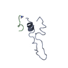
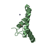
 Movie
Movie Controller


+ Open data

- Basic information
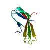
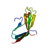
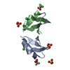
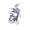
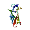
| Entry | Database: PDB / ID: 5mj1 | |||||||||
|---|---|---|---|---|---|---|---|---|---|---|
| Title | Extracellular domain of human CD83 - rhombohedral crystal form | |||||||||
 Components Components | CD83 antigen | |||||||||
 Keywords Keywords | IMMUNE SYSTEM / Dendritic cell / receptor / immunoglobulin | |||||||||
| Function / homology |  Function and homology information Function and homology informationpositive regulation of CD4-positive, alpha-beta T cell differentiation / negative regulation of interleukin-4 production / CD4-positive, alpha-beta T cell differentiation / humoral immune response / positive regulation of interleukin-10 production / positive regulation of interleukin-2 production / defense response / external side of plasma membrane / signal transduction / plasma membrane Similarity search - Function | |||||||||
| Biological species |  Homo sapiens (human) Homo sapiens (human) | |||||||||
| Method |  X-RAY DIFFRACTION / SYNCHROTRON / MOLECULAR REPLACEMENT / Resolution: 1.8 Å X-RAY DIFFRACTION / SYNCHROTRON / MOLECULAR REPLACEMENT / Resolution: 1.8 Å | |||||||||
 Authors Authors | Klingl, S. / Egerer-Sieber, C. / Schmid, B. / Weiler, S. / Muller, Y.A. | |||||||||
| Funding support |  Germany, 1items Germany, 1items
| |||||||||
 Citation Citation | Journal: J. Mol. Biol. / Year: 2017 Title: Crystal Structure of the Extracellular Domain of the Human Dendritic Cell Surface Marker CD83. Authors: Heilingloh, C.S. / Klingl, S. / Egerer-Sieber, C. / Schmid, B. / Weiler, S. / Muhl-Zurbes, P. / Hofmann, J. / Stump, J.D. / Sticht, H. / Kummer, M. / Steinkasserer, A. / Muller, Y.A. | |||||||||
| History |
|
- Structure visualization
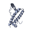
| Structure viewer | Molecule: MolmilJmol/JSmol |
|---|
- Downloads & links
-Download
| PDBx/mmCIF format | 5mj1.cif.gz | 51.3 KB | Display | PDBx/mmCIF format |
|---|---|---|---|---|
| PDB format | pdb5mj1.ent.gz | 35.9 KB | Display | PDB format |
| PDBx/mmJSON format | 5mj1.json.gz | Tree view | PDBx/mmJSON format | |
| Others |  Other downloads Other downloads |
-Validation report
| Summary document | 5mj1_validation.pdf.gz | 421.6 KB | Display | wwPDB validaton report |
|---|---|---|---|---|
| Full document | 5mj1_full_validation.pdf.gz | 422.3 KB | Display | |
| Data in XML | 5mj1_validation.xml.gz | 7.2 KB | Display | |
| Data in CIF | 5mj1_validation.cif.gz | 8.6 KB | Display | |
| Arichive directory | https://data.pdbj.org/pub/pdb/validation_reports/mj/5mj1ftp://data.pdbj.org/pub/pdb/validation_reports/mj/5mj1 | HTTPS FTP |
-Related structure data


-Links
 PDBj
PDBj- Assembly
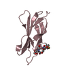
| Deposited unit | 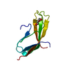
| |||||||||
|---|---|---|---|---|---|---|---|---|---|---|
| 1 |
| |||||||||
| Unit cell |
| |||||||||
| Components on special symmetry positions |
|
-Components
| #1: Protein | Mass: 12766.979 Da / Num. of mol.: 1 / Mutation: C27S, C100S, C129S Source method: isolated from a genetically manipulated source Details: The first four residues (GSPG) are non-native residues of the linker which remain after the GST-tag was cleaved off. The third residue (P) was modeled as alanine due to missing electron ...Details: The first four residues (GSPG) are non-native residues of the linker which remain after the GST-tag was cleaved off. The third residue (P) was modeled as alanine due to missing electron density. The first and last two residues as well as the central region were not visible in the electron density maps. Source: (gene. exp.) Homo sapiens (human) / Cell: Dendritic cells / Gene: CD83 / Plasmid: pGEX-2T / Production host: Escherichia coli 'BL21-Gold(DE3)pLysS AG' / References: UniProt: Q01151 |
|---|---|
| #2: Chemical | ChemComp-PEG /   Mass: 106.120 Da / Num. of mol.: 1 / Source method: obtained synthetically / Formula: C4H10O3 Mass: 106.120 Da / Num. of mol.: 1 / Source method: obtained synthetically / Formula: C4H10O3 |
| #3: Water | ChemComp-HOH /  Mass: 18.015 Da / Num. of mol.: 58 / Source method: isolated from a natural source / Formula: H2O Mass: 18.015 Da / Num. of mol.: 58 / Source method: isolated from a natural source / Formula: H2O |
| Has protein modification | Y |
-Experimental details
-Experiment
| Experiment | Method: X-RAY DIFFRACTION / Number of used crystals: 1 |
|---|
- Sample preparation
| Crystal | Density Matthews: 2 Å3/Da / Density % sol: 38.64 % |
|---|---|
| Crystal grow | Temperature: 293 K / Method: vapor diffusion, sitting drop Details: 0.2 microliter protein (39 mg/ml in ultrapure water) mixed with 0.4 microliter reservoir solution (0.2 M DL-malic acid (pH 7.0), 20% w/v PEG 3350) was equilibrated against 70 microliter of ...Details: 0.2 microliter protein (39 mg/ml in ultrapure water) mixed with 0.4 microliter reservoir solution (0.2 M DL-malic acid (pH 7.0), 20% w/v PEG 3350) was equilibrated against 70 microliter of reservoir solution. Crystals appeared after ~ 13 months |
-Data collection
| Diffraction | Mean temperature: 100 K |
|---|---|
| Diffraction source | Source: SYNCHROTRON / Site: BESSY / Beamline: 14.1 / Wavelength: 0.918409 Å |
| Detector | Type: DECTRIS PILATUS 6M / Detector: PIXEL / Date: Mar 27, 2014 |
| Radiation | Protocol: SINGLE WAVELENGTH / Monochromatic (M) / Laue (L): M / Scattering type: x-ray |
| Radiation wavelength | Wavelength: 0.918409 Å / Relative weight: 1 |
| Reflection | Resolution: 1.8→31.4 Å / Num. obs: 9772 / % possible obs: 99.8 % / Redundancy: 5.4 % / Biso Wilson estimate: 33.7 Å2 / Rmerge(I) obs: 0.053 / Net I/σ(I): 21 |
| Reflection shell | Resolution: 1.8→1.91 Å / Redundancy: 5.5 % / Rmerge(I) obs: 0.74 / Mean I/σ(I) obs: 2.2 / % possible all: 99.7 |
- Processing
| Software |
| ||||||||||||||||||||||||||||||||||||||||||||||||||||||||||||||||||||||||||||||||||||||||||||||||||||||||||||||||||||||||||||||||||||||||||||||||||||||||||||||||||||||||||||||||||||||||||||||||||||||||
|---|---|---|---|---|---|---|---|---|---|---|---|---|---|---|---|---|---|---|---|---|---|---|---|---|---|---|---|---|---|---|---|---|---|---|---|---|---|---|---|---|---|---|---|---|---|---|---|---|---|---|---|---|---|---|---|---|---|---|---|---|---|---|---|---|---|---|---|---|---|---|---|---|---|---|---|---|---|---|---|---|---|---|---|---|---|---|---|---|---|---|---|---|---|---|---|---|---|---|---|---|---|---|---|---|---|---|---|---|---|---|---|---|---|---|---|---|---|---|---|---|---|---|---|---|---|---|---|---|---|---|---|---|---|---|---|---|---|---|---|---|---|---|---|---|---|---|---|---|---|---|---|---|---|---|---|---|---|---|---|---|---|---|---|---|---|---|---|---|---|---|---|---|---|---|---|---|---|---|---|---|---|---|---|---|---|---|---|---|---|---|---|---|---|---|---|---|---|---|---|---|---|
| Refinement | Method to determine structure: MOLECULAR REPLACEMENT / Resolution: 1.8→31.4 Å / SU ML: 0.21 / Cross valid method: FREE R-VALUE / σ(F): 1.36 / Phase error: 27.7
| ||||||||||||||||||||||||||||||||||||||||||||||||||||||||||||||||||||||||||||||||||||||||||||||||||||||||||||||||||||||||||||||||||||||||||||||||||||||||||||||||||||||||||||||||||||||||||||||||||||||||
| Solvent computation | Shrinkage radii: 0.9 Å / VDW probe radii: 1.11 Å | ||||||||||||||||||||||||||||||||||||||||||||||||||||||||||||||||||||||||||||||||||||||||||||||||||||||||||||||||||||||||||||||||||||||||||||||||||||||||||||||||||||||||||||||||||||||||||||||||||||||||
| Refinement step | Cycle: LAST / Resolution: 1.8→31.4 Å
| ||||||||||||||||||||||||||||||||||||||||||||||||||||||||||||||||||||||||||||||||||||||||||||||||||||||||||||||||||||||||||||||||||||||||||||||||||||||||||||||||||||||||||||||||||||||||||||||||||||||||
| Refine LS restraints |
| ||||||||||||||||||||||||||||||||||||||||||||||||||||||||||||||||||||||||||||||||||||||||||||||||||||||||||||||||||||||||||||||||||||||||||||||||||||||||||||||||||||||||||||||||||||||||||||||||||||||||
| LS refinement shell |
| ||||||||||||||||||||||||||||||||||||||||||||||||||||||||||||||||||||||||||||||||||||||||||||||||||||||||||||||||||||||||||||||||||||||||||||||||||||||||||||||||||||||||||||||||||||||||||||||||||||||||
| Refinement TLS params. | Method: refined / Refine-ID: X-RAY DIFFRACTION
| ||||||||||||||||||||||||||||||||||||||||||||||||||||||||||||||||||||||||||||||||||||||||||||||||||||||||||||||||||||||||||||||||||||||||||||||||||||||||||||||||||||||||||||||||||||||||||||||||||||||||
| Refinement TLS group |
|
