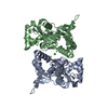
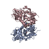
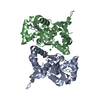
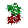
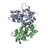
Mass: 29108.453 Da / Num. of mol.: 2 / Fragment: UNP residues 445-559,UNP residues 682-820 Source method: isolated from a genetically manipulated source Details: THE PROTEIN CRYSTALLIZED IS THE EXTRACELLULAR LIGAND BINDING DOMAIN OF GLUK1. TRANSMEMBRANE REGIONS WERE GENETICALLY REMOVED AND REPLACED WITH A GLY-THR LINKER (RESIDUES 545 AND 546 OF THE ...Details: THE PROTEIN CRYSTALLIZED IS THE EXTRACELLULAR LIGAND BINDING DOMAIN OF GLUK1. TRANSMEMBRANE REGIONS WERE GENETICALLY REMOVED AND REPLACED WITH A GLY-THR LINKER (RESIDUES 545 AND 546 OF THE STRUCTURE). THEREFORE, THE SEQUENCE MATCHES DISCONTINUOUSLY WITH THE REFERENCE DATABASE (430-544, 667-805). RESIDUE 429 IS A REMNANT FROM CLONING. Source: (gene. exp.) Rattus norvegicus (Norway rat) / Gene: Grik1, Glur5 / Plasmid: PET28A / Production host: Escherichia coli (E. coli) / Variant (production host): ORIGAMI 2 / References: UniProt: P22756
Mass: 18.015 Da / Num. of mol.: 324 / Source method: isolated from a natural source / Formula: H2O
-
Details
Has protein modification
Y
Sequence details
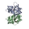
THE SEQUENCE DATABASE IS P22756-2, ISOFORM GLUR5-2. THE PROTEIN CRYSTALLIZED IS THE EXTRACELLULAR ...THE SEQUENCE DATABASE IS P22756-2, ISOFORM GLUR5-2. THE PROTEIN CRYSTALLIZED IS THE EXTRACELLULAR LIGAND-BINDING DOMAIN OF GLUK1. TRANSMEMBRANE REGIONS WERE GENETICALLY REMOVED AND REPLACED WITH A GLY-THR LINKER. THERE IS A SEQUENCE CONFLICT AT RESIDUE 462 OF THE CRYSTALLIZED PROTEIN DUE TO DIFFERENCES IN DATABASE SEQUENCE (SEE GENBANK ACCESION NO.AAA02874).
-
Experimental details
-
Experiment
Experiment
Method: X-RAY DIFFRACTION / Number of used crystals: 1
-
Sample preparation
Crystal
Density Matthews: 2.4 Å3/Da / Density % sol: 48.73 %
Crystal grow
Temperature: 279 K / Method: vapor diffusion, hanging drop / pH: 4.5 Details: 16 % PEG4000, 0.2 M lithium-sulfate, 0.1 M phosphate-citrate pH 4.5
-
Data collection
Diffraction
Mean temperature: 100 K
Diffraction source
Source: SYNCHROTRON / Site: MAX II / Beamline: I911-3 / Wavelength: 0.97879 Å
 Movie
Movie Controller

 Yorodumi
 Open data
 Basic information
 Components
 Keywords
 Function and homology information
 X-RAY DIFFRACTION /
  Authors
 Citation
 Structure visualization
 Downloads & links
 Other downloads


 PDBj
PDBj

 Assembly
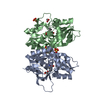


 Mass: 240.279 Da / Num. of mol.: 2 / Source method: obtained synthetically / Formula: C10H12N2O3S
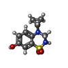
 Mass: 213.230 Da / Num. of mol.: 2 / Source method: obtained synthetically / Formula: C10H15NO4 / Comment: neurotransmitter, agonist*YM
 Mass: 35.453 Da / Num. of mol.: 1 / Source method: obtained synthetically / Formula: Cl
 Mass: 96.063 Da / Num. of mol.: 3 / Source method: obtained synthetically / Formula: SO4
 Mass: 92.094 Da / Num. of mol.: 1 / Source method: obtained synthetically / Formula: C3H8O3
 Mass: 59.044 Da / Num. of mol.: 1 / Source method: obtained synthetically / Formula: C2H3O2
 Sample preparation
 / Beamline: I911-3 / Wavelength: 0.97879 Å
 Processing