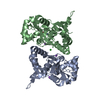
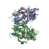
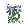
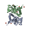
Mass: 29425.621 Da / Num. of mol.: 2 / Fragment: residues 429-544, 667-806 / Mutation: I473H, K525E, K696R, I780L, Q784K Source method: isolated from a genetically manipulated source Details: The I442H K494E K665R I749L and Q753K mutations were created intentionally. The 1st residue is a vector encoded affinity tag fragment. Residues 398-513 and 636 -775 are coupled by a ...Details: The I442H K494E K665R I749L and Q753K mutations were created intentionally. The 1st residue is a vector encoded affinity tag fragment. Residues 398-513 and 636 -775 are coupled by a synthetic GT peptide. The numbering is for the mature prot ein after cleavage of the 31 AA signal peptide. Source: (gene. exp.) Rattus norvegicus (Norway rat) / Gene: GriK2 / Plasmid: pET22B modified / Production host: Escherichia coli (E. coli) / Strain (production host): OrigamiB (DE3) / References: UniProt: P42260
Resolution: 1.32→1.37 Å / Redundancy: 3.4 % / Rmerge(I) obs: 0.369 / Mean I/σ(I) obs: 3.4 / % possible all: 99.2
-
Processing
Software
Name
Version
Classification
HKL-2000
datacollection
PHENIX
(phenix.refine)
refinement
HKL-2000
datareduction
HKL-2000
datascaling
Refinement
Method to determine structure: FOURIER SYNTHESIS / Resolution: 1.321→34.516 Å / SU ML: 0.17 / Cross valid method: THROUGHOUT / σ(F): 0 / σ(I): 0 / Stereochemistry target values: ML Details: Refinement was started with Refmac_5.2. The final rounds of refinement were performed with phenix and included occupancy refinement for ions, and for residues with alternative conformations.
Rfactor
Num. reflection
% reflection
Selection details
Rfree
0.1789
6231
5.01 %
RANDOM
Rwork
0.1561
-
-
-
all
0.1573
124266
-
-
obs
0.1573
124266
99.74 %
-
Solvent computation
Shrinkage radii: 0.9 Å / VDW probe radii: 1.11 Å / Solvent model: FLAT BULK SOLVENT MODEL / Bsol: 52.808 Å2 / ksol: 0.414 e/Å3
Refinement step
Cycle: LAST / Resolution: 1.321→34.516 Å
Protein
Nucleic acid
Ligand
Solvent
Total
Num. atoms
8753
0
39
618
9410
Refine LS restraints
Refine-ID
Type
Dev ideal
Number
X-RAY DIFFRACTION
f_bond_d
0.01
8905
X-RAY DIFFRACTION
f_angle_d
1.114
16188
X-RAY DIFFRACTION
f_chiral_restr
0.089
677
X-RAY DIFFRACTION
f_plane_restr
0.006
1368
X-RAY DIFFRACTION
f_dihedral_angle_d
14.865
2307
LS refinement shell
Resolution (Å)
Rfactor Rfree
Num. reflection Rfree
Rfactor Rwork
Num. reflection Rwork
Refine-ID
% reflection obs (%)
1.321-1.336
0.2291
214
0.2219
3767
X-RAY DIFFRACTION
95
1.336-1.3518
0.233
233
0.2112
3858
X-RAY DIFFRACTION
99
1.3518-1.3682
0.2251
206
0.2095
3962
X-RAY DIFFRACTION
100
1.3682-1.3856
0.2443
204
0.1994
3903
X-RAY DIFFRACTION
100
1.3856-1.4038
0.1855
196
0.1917
3980
X-RAY DIFFRACTION
100
1.4038-1.423
0.2093
206
0.1824
3894
X-RAY DIFFRACTION
100
1.423-1.4434
0.1964
204
0.1747
3941
X-RAY DIFFRACTION
100
1.4434-1.4649
0.2095
181
0.1668
3947
X-RAY DIFFRACTION
100
1.4649-1.4878
0.1736
227
0.1588
3935
X-RAY DIFFRACTION
100
1.4878-1.5122
0.1774
196
0.152
3898
X-RAY DIFFRACTION
100
1.5122-1.5383
0.1733
203
0.1506
4006
X-RAY DIFFRACTION
100
1.5383-1.5662
0.1788
188
0.1462
3897
X-RAY DIFFRACTION
100
1.5662-1.5964
0.1662
203
0.1461
3955
X-RAY DIFFRACTION
100
1.5964-1.6289
0.1664
202
0.141
3937
X-RAY DIFFRACTION
100
1.6289-1.6644
0.1836
202
0.1403
3937
X-RAY DIFFRACTION
100
1.6644-1.7031
0.1662
195
0.1477
3955
X-RAY DIFFRACTION
100
1.7031-1.7457
0.1754
204
0.1455
3946
X-RAY DIFFRACTION
100
1.7457-1.7929
0.1779
210
0.1464
3929
X-RAY DIFFRACTION
100
1.7929-1.8456
0.1733
213
0.1439
3939
X-RAY DIFFRACTION
100
1.8456-1.9052
0.16
212
0.1445
3926
X-RAY DIFFRACTION
100
1.9052-1.9733
0.1712
208
0.1442
3967
X-RAY DIFFRACTION
100
1.9733-2.0523
0.1544
216
0.1315
3920
X-RAY DIFFRACTION
100
2.0523-2.1457
0.1602
205
0.1279
3945
X-RAY DIFFRACTION
100
2.1457-2.2588
0.1457
207
0.1286
3951
X-RAY DIFFRACTION
100
2.2588-2.4002
0.1595
220
0.1363
3935
X-RAY DIFFRACTION
100
2.4002-2.5855
0.1728
214
0.1436
3940
X-RAY DIFFRACTION
100
2.5855-2.8456
0.184
227
0.1497
3949
X-RAY DIFFRACTION
100
2.8456-3.2571
0.1713
213
0.1471
3961
X-RAY DIFFRACTION
100
3.2571-4.1025
0.1509
225
0.1389
3942
X-RAY DIFFRACTION
100
4.1025-34.5283
0.1792
197
0.1574
4013
X-RAY DIFFRACTION
99
Refinement TLS params.
Method: refined / Refine-ID: X-RAY DIFFRACTION
ID
L11 (°2)
L12 (°2)
L13 (°2)
L22 (°2)
L23 (°2)
L33 (°2)
S11 (Å °)
S12 (Å °)
S13 (Å °)
S21 (Å °)
S22 (Å °)
S23 (Å °)
S31 (Å °)
S32 (Å °)
S33 (Å °)
T11 (Å2)
T12 (Å2)
T13 (Å2)
T22 (Å2)
T23 (Å2)
T33 (Å2)
Origin x (Å)
Origin y (Å)
Origin z (Å)
1
0.7529
0.4013
-0.004
0.97
-0.2205
0.8205
-0.0625
0.0961
0.1111
-0.2486
0.0565
0.0059
-0.0742
0.0152
-0.0044
0.1556
-0.0054
0.0136
0.0971
0.0284
0.0984
14.744
29.7692
-5.5416
2
0.6112
0.2795
0.3547
0.9629
0.2723
0.5364
0.0131
-0.0052
0.0759
0.0535
-0.0095
-0.055
-0.0063
0.0265
-0.0407
0.0619
-0.0033
0.0129
0.0481
0
0.0612
20.628
31.2667
14.6013
3
0.1079
0.0379
-0.0754
0.6765
-0.1037
0.3672
-0.0116
0.1655
-0.0439
-0.1237
0.0331
-0.1353
0.0769
0.0761
-0.0284
0.1323
0.008
0.0254
0.1227
-0.0017
0.1205
19.6622
14.1161
-1.5707
4
0.9294
-0.0826
-0.0259
0.7475
0.0741
0.7371
0.0208
-0.066
-0.0579
0.0139
0.0067
0.0734
0.0226
-0.046
-0.0275
0.066
-0.0073
-0.0087
0.0836
0.0101
0.0784
4.9763
0.8793
16.695
5
1.1698
-0.3154
-0.0664
0.5402
-0.0152
0.8043
0.0398
-0.0564
-0.0794
0.007
-0.0265
-0.0775
0.0637
0.0783
0.0068
0.0111
0.006
-0.0048
0.0296
-0.001
0.0307
27.4244
-4.8433
11.2925
6
0.2798
-0.0065
-0.1162
0.5236
-0.2452
0.2704
0.0288
-0.0572
0.0798
0.0446
0.027
0.0165
-0.088
0.016
-0.0497
0.0944
-0.0025
0.0042
0.0929
-0.0127
0.0903
8.0508
13.1899
17.318
Refinement TLS group
ID
Refine-ID
Refine TLS-ID
Selection details
1
X-RAY DIFFRACTION
1
chainAand (resid3:86)
2
X-RAY DIFFRACTION
2
chainAand (resid87:226)
3
X-RAY DIFFRACTION
3
chainAand (resid227:253)
4
X-RAY DIFFRACTION
4
chainBand (resid2:108)
5
X-RAY DIFFRACTION
5
chainBand (resid109:216)
6
X-RAY DIFFRACTION
6
chainBand (resid217:253)
+
About Yorodumi
-
News
-
Feb 9, 2022. New format data for meta-information of EMDB entries
New format data for meta-information of EMDB entries
Version 3 of the EMDB header file is now the official format.
The previous official version 1.9 will be removed from the archive.
In the structure databanks used in Yorodumi, some data are registered as the other names, "COVID-19 virus" and "2019-nCoV". Here are the details of the virus and the list of structure data.
Jan 31, 2019. EMDB accession codes are about to change! (news from PDBe EMDB page)
EMDB accession codes are about to change! (news from PDBe EMDB page)
The allocation of 4 digits for EMDB accession codes will soon come to an end. Whilst these codes will remain in use, new EMDB accession codes will include an additional digit and will expand incrementally as the available range of codes is exhausted. The current 4-digit format prefixed with “EMD-” (i.e. EMD-XXXX) will advance to a 5-digit format (i.e. EMD-XXXXX), and so on. It is currently estimated that the 4-digit codes will be depleted around Spring 2019, at which point the 5-digit format will come into force.
The EM Navigator/Yorodumi systems omit the EMD- prefix.
Related info.:Q: What is EMD? / ID/Accession-code notation in Yorodumi/EM Navigator
Yorodumi is a browser for structure data from EMDB, PDB, SASBDB, etc.
This page is also the successor to EM Navigator detail page, and also detail information page/front-end page for Omokage search.
The word "yorodu" (or yorozu) is an old Japanese word meaning "ten thousand". "mi" (miru) is to see.
Related info.:EMDB / PDB / SASBDB / Comparison of 3 databanks / Yorodumi Search / Aug 31, 2016. New EM Navigator & Yorodumi / Yorodumi Papers / Jmol/JSmol / Function and homology information / Changes in new EM Navigator and Yorodumi
 Movie
Movie Controller

 Yorodumi
 Open data
 Basic information
 Components
 Keywords
 Function and homology information
 X-RAY DIFFRACTION /
  Authors
 Citation
 Structure visualization
 Downloads & links
 Other downloads
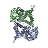


 PDBj
PDBj


 Assembly
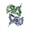


 Type: L-peptide linking / Mass: 147.129 Da / Num. of mol.: 2 / Source method: obtained synthetically / Formula: C5H9NO4
 Mass: 35.453 Da / Num. of mol.: 2 / Source method: obtained synthetically / Formula: Cl
 Mass: 22.990 Da / Num. of mol.: 4 / Source method: obtained synthetically / Formula: Na
 Mass: 18.015 Da / Num. of mol.: 618 / Source method: isolated from a natural source / Formula: H2O
 Sample preparation
 / Beamline: 22-ID / Wavelength: 1 Å
 Processing