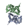
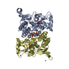
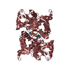
- PDB-3g3k: Crystal structure of the GluR6 ligand binding domain dimer I442H ... -
+
Open data
ID or keywords:
Loading...
-
Basic information
Entry
Database: PDB / ID: 3g3k
Title
Crystal structure of the GluR6 ligand binding domain dimer I442H K494E K665R I749L Q753K E757Q mutant with glutamate and NaCl at 1.24 Angstrom resolution
Components
Glutamate receptor, ionotropic kainate 2
Keywords
MEMBRANE PROTEIN / Cell junction / Cell membrane / Glycoprotein / Ion transport / Ionic channel / Membrane / Postsynaptic cell membrane / Receptor / RNA editing / Synapse / Transmembrane / Transport
Function / homology
Function and homology information
mossy fiber rosette / detection of cold stimulus involved in thermoception / Activation of Na-permeable kainate receptors / regulation of short-term neuronal synaptic plasticity / Activation of Ca-permeable Kainate Receptor / kainate selective glutamate receptor complex / ubiquitin conjugating enzyme binding / glutamate receptor activity / negative regulation of synaptic transmission, glutamatergic / regulation of JNK cascade ...mossy fiber rosette / detection of cold stimulus involved in thermoception / Activation of Na-permeable kainate receptors / regulation of short-term neuronal synaptic plasticity / Activation of Ca-permeable Kainate Receptor / kainate selective glutamate receptor complex / ubiquitin conjugating enzyme binding / glutamate receptor activity / negative regulation of synaptic transmission, glutamatergic / regulation of JNK cascade / inhibitory postsynaptic potential / receptor clustering / kainate selective glutamate receptor activity / extracellularly glutamate-gated ion channel activity / modulation of excitatory postsynaptic potential / ionotropic glutamate receptor complex / behavioral fear response / positive regulation of synaptic transmission / neuronal action potential / glutamate-gated receptor activity / glutamate-gated calcium ion channel activity / dendrite cytoplasm / ligand-gated monoatomic ion channel activity involved in regulation of presynaptic membrane potential / presynaptic modulation of chemical synaptic transmission / excitatory postsynaptic potential / hippocampal mossy fiber to CA3 synapse / SNARE binding / PDZ domain binding / regulation of long-term neuronal synaptic plasticity / synaptic transmission, glutamatergic / regulation of membrane potential / transmitter-gated monoatomic ion channel activity involved in regulation of postsynaptic membrane potential / intracellular protein transport / postsynaptic density membrane / modulation of chemical synaptic transmission / intracellular calcium ion homeostasis / terminal bouton / positive regulation of neuron apoptotic process / neuron apoptotic process / presynaptic membrane / scaffold protein binding / chemical synaptic transmission / negative regulation of neuron apoptotic process / perikaryon / postsynaptic membrane / postsynaptic density / axon / neuronal cell body / ubiquitin protein ligase binding / dendrite / synapse / glutamatergic synapse / membrane / identical protein binding / plasma membrane Similarity search - Function
Ionotropic glutamate receptor, metazoa / Ligated ion channel L-glutamate- and glycine-binding site / Ligand-gated ion channel / Ionotropic glutamate receptor, L-glutamate and glycine-binding domain / Ligated ion channel L-glutamate- and glycine-binding site / : / Ionotropic glutamate receptor / Eukaryotic homologues of bacterial periplasmic substrate binding proteins. / Periplasmic binding protein-like II / Receptor, ligand binding region ...Ionotropic glutamate receptor, metazoa / Ligated ion channel L-glutamate- and glycine-binding site / Ligand-gated ion channel / Ionotropic glutamate receptor, L-glutamate and glycine-binding domain / Ligated ion channel L-glutamate- and glycine-binding site / : / Ionotropic glutamate receptor / Eukaryotic homologues of bacterial periplasmic substrate binding proteins. / Periplasmic binding protein-like II / Receptor, ligand binding region / Receptor family ligand binding region / D-Maltodextrin-Binding Protein; domain 2 / Periplasmic binding protein-like I / 3-Layer(aba) Sandwich / Alpha Beta Similarity search - Domain/homology
Mass: 29424.637 Da / Num. of mol.: 2 / Fragment: residues 429-544, 667-806 / Mutation: I473H, K525E, K696R, I780L, Q784K, E788Q Source method: isolated from a genetically manipulated source Details: The I442H K494E K665R I749L Q753K and E757Q mutations were created intentionall y. The 1st residue is a vector encoded affinity tag fragment. Residues 398-513 a nd 636-775 are coupled by a ...Details: The I442H K494E K665R I749L Q753K and E757Q mutations were created intentionall y. The 1st residue is a vector encoded affinity tag fragment. Residues 398-513 a nd 636-775 are coupled by a synthetic GT peptide. The numbering is for the matur e protein after cleavage of the 31 AA signal peptide. Source: (gene. exp.) Rattus norvegicus (Norway rat) / Gene: GriK2 / Plasmid: pET22B modified / Production host: Escherichia coli (E. coli) / Strain (production host): OrigamiB (DE3) / References: UniProt: P42260
Resolution: 1.24→1.28 Å / Redundancy: 2.8 % / Rmerge(I) obs: 0.282 / Mean I/σ(I) obs: 3.39 / % possible all: 86.5
-
Processing
Software
Name
Version
Classification
HKL-2000
datacollection
PHENIX
(phenix.refine)
refinement
HKL-2000
datareduction
HKL-2000
datascaling
Refinement
Method to determine structure: FOURIER SYNTHESIS / Resolution: 1.24→35.898 Å / SU ML: 0.14 / Isotropic thermal model: ISOTROPIC AND TLS / Cross valid method: THROUGHOUT / σ(F): 0 / σ(I): 0 / Stereochemistry target values: ML Details: Refinement was started with Refmac_5.2. The final rounds of refinement were performed with phenix and included occupancy refinement for ions, and for residues with alternative conformations.
Rfactor
Num. reflection
% reflection
Selection details
Rfree
0.165
7489
5.03 %
RANDOM
Rwork
0.1459
-
-
-
all
0.1469
149020
-
-
obs
0.1469
149020
98.18 %
-
Solvent computation
Shrinkage radii: 0.9 Å / VDW probe radii: 1.11 Å / Solvent model: FLAT BULK SOLVENT MODEL / Bsol: 41.237 Å2 / ksol: 0.393 e/Å3
Refinement step
Cycle: LAST / Resolution: 1.24→35.898 Å
Protein
Nucleic acid
Ligand
Solvent
Total
Num. atoms
9668
0
41
752
10461
Refine LS restraints
Refine-ID
Type
Dev ideal
Number
X-RAY DIFFRACTION
f_bond_d
0.017
9916
X-RAY DIFFRACTION
f_angle_d
1.388
18183
X-RAY DIFFRACTION
f_chiral_restr
0.126
734
X-RAY DIFFRACTION
f_plane_restr
0.008
1518
X-RAY DIFFRACTION
f_dihedral_angle_d
14.97
2651
LS refinement shell
Resolution (Å)
Rfactor Rfree
Num. reflection Rfree
Rfactor Rwork
Num. reflection Rwork
Refine-ID
% reflection obs (%)
1.2403-1.2544
0.2387
172
0.2236
3189
X-RAY DIFFRACTION
67
1.2544-1.2691
0.2269
206
0.2164
4077
X-RAY DIFFRACTION
85
1.2691-1.2846
0.2228
243
0.1969
4547
X-RAY DIFFRACTION
95
1.2846-1.3009
0.1947
283
0.1834
4752
X-RAY DIFFRACTION
100
1.3009-1.318
0.1936
236
0.1723
4824
X-RAY DIFFRACTION
100
1.318-1.336
0.193
266
0.1739
4765
X-RAY DIFFRACTION
100
1.336-1.3551
0.187
294
0.1624
4761
X-RAY DIFFRACTION
100
1.3551-1.3754
0.1756
235
0.1587
4828
X-RAY DIFFRACTION
100
1.3754-1.3969
0.1758
253
0.1537
4747
X-RAY DIFFRACTION
100
1.3969-1.4198
0.1621
260
0.1454
4832
X-RAY DIFFRACTION
100
1.4198-1.4442
0.1466
235
0.1485
4780
X-RAY DIFFRACTION
100
1.4442-1.4705
0.1704
230
0.1433
4850
X-RAY DIFFRACTION
100
1.4705-1.4988
0.1825
261
0.1395
4822
X-RAY DIFFRACTION
100
1.4988-1.5294
0.1521
246
0.1321
4781
X-RAY DIFFRACTION
100
1.5294-1.5626
0.1659
233
0.1327
4795
X-RAY DIFFRACTION
100
1.5626-1.599
0.1499
257
0.1338
4827
X-RAY DIFFRACTION
100
1.599-1.639
0.1547
237
0.1321
4827
X-RAY DIFFRACTION
100
1.639-1.6833
0.1679
256
0.1383
4791
X-RAY DIFFRACTION
100
1.6833-1.7328
0.1592
225
0.1383
4842
X-RAY DIFFRACTION
100
1.7328-1.7887
0.1664
263
0.1374
4783
X-RAY DIFFRACTION
100
1.7887-1.8527
0.1598
260
0.14
4790
X-RAY DIFFRACTION
100
1.8527-1.9268
0.1698
269
0.1387
4774
X-RAY DIFFRACTION
100
1.9268-2.0145
0.1463
245
0.1346
4816
X-RAY DIFFRACTION
100
2.0145-2.1207
0.1517
258
0.126
4807
X-RAY DIFFRACTION
100
2.1207-2.2536
0.1404
248
0.1235
4781
X-RAY DIFFRACTION
100
2.2536-2.4275
0.1435
274
0.13
4805
X-RAY DIFFRACTION
100
2.4275-2.6718
0.1627
261
0.138
4784
X-RAY DIFFRACTION
100
2.6718-3.0582
0.1775
261
0.1495
4847
X-RAY DIFFRACTION
100
3.0582-3.8523
0.1486
278
0.1345
4800
X-RAY DIFFRACTION
100
3.8523-35.9133
0.1543
244
0.1446
4907
X-RAY DIFFRACTION
100
Refinement TLS params.
Method: refined / Refine-ID: X-RAY DIFFRACTION
ID
L11 (°2)
L12 (°2)
L13 (°2)
L22 (°2)
L23 (°2)
L33 (°2)
S11 (Å °)
S12 (Å °)
S13 (Å °)
S21 (Å °)
S22 (Å °)
S23 (Å °)
S31 (Å °)
S32 (Å °)
S33 (Å °)
T11 (Å2)
T12 (Å2)
T13 (Å2)
T22 (Å2)
T23 (Å2)
T33 (Å2)
Origin x (Å)
Origin y (Å)
Origin z (Å)
1
0.466
0.2035
-0.0187
0.6649
-0.0479
0.4091
-0.0277
0.0555
0.0728
-0.134
0.0431
0.0227
-0.0377
-0.0062
-0.0088
0.1134
0.0003
0.0087
0.0772
0.0224
0.0819
14.1805
28.4575
-3.1756
2
0.4031
0.087
0.1965
0.4995
0.1996
0.5105
0.0155
-0.0013
0.0795
0.0391
-0.0223
-0.0809
-0.0079
0.0362
-0.0023
0.092
-0.0061
0.0109
0.0757
-0.0111
0.1046
24.1391
33.8197
17.0593
3
0.046
0.0151
0.04
0.3557
-0.0258
0.2163
0.005
0.0158
0.0181
-0.1113
0.0198
-0.0735
0.0391
0.0228
0.002
0.1008
0.0039
0.0153
0.0852
0.0019
0.0882
19.3513
15.6481
-0.7787
4
0.7035
-0.0363
-0.0438
0.4691
0.087
0.4336
0.0066
-0.0378
-0.0595
0.0174
0.0154
0.0576
0.0194
-0.034
0.0162
0.0509
-0.0038
-0.0088
0.0643
0.0094
0.0595
5.1476
0.2671
16.6071
5
0.6618
-0.159
-0.0835
0.2366
-0.0878
0.4976
0.0229
-0.0236
-0.0761
-0.011
-0.0218
-0.0429
0.0333
0.0341
0
0.0408
0.0075
-0.0067
0.054
-0.0042
0.0561
27.4758
-4.9064
11.7231
6
0.2416
-0.0654
-0.1476
0.3038
-0.0943
0.2002
0.0392
-0.0361
0.0677
0.0608
0.0203
0.0014
-0.0321
0.0167
0.0225
0.0814
-0.0019
-0.0017
0.0769
-0.009
0.073
9.6335
13.075
19.1552
Refinement TLS group
ID
Refine-ID
Refine TLS-ID
Selection details
1
X-RAY DIFFRACTION
1
chainAand (resid3:107)
2
X-RAY DIFFRACTION
2
chainAand (resid108:216)
3
X-RAY DIFFRACTION
3
chainAand (resid217:258)
4
X-RAY DIFFRACTION
4
chainBand (resid2:108)
5
X-RAY DIFFRACTION
5
chainBand (resid109:216)
6
X-RAY DIFFRACTION
6
chainBand (resid217:257)
+
About Yorodumi
-
News
-
Feb 9, 2022. New format data for meta-information of EMDB entries
New format data for meta-information of EMDB entries
Version 3 of the EMDB header file is now the official format.
The previous official version 1.9 will be removed from the archive.
In the structure databanks used in Yorodumi, some data are registered as the other names, "COVID-19 virus" and "2019-nCoV". Here are the details of the virus and the list of structure data.
Jan 31, 2019. EMDB accession codes are about to change! (news from PDBe EMDB page)
EMDB accession codes are about to change! (news from PDBe EMDB page)
The allocation of 4 digits for EMDB accession codes will soon come to an end. Whilst these codes will remain in use, new EMDB accession codes will include an additional digit and will expand incrementally as the available range of codes is exhausted. The current 4-digit format prefixed with “EMD-” (i.e. EMD-XXXX) will advance to a 5-digit format (i.e. EMD-XXXXX), and so on. It is currently estimated that the 4-digit codes will be depleted around Spring 2019, at which point the 5-digit format will come into force.
The EM Navigator/Yorodumi systems omit the EMD- prefix.
Related info.:Q: What is EMD? / ID/Accession-code notation in Yorodumi/EM Navigator
Yorodumi is a browser for structure data from EMDB, PDB, SASBDB, etc.
This page is also the successor to EM Navigator detail page, and also detail information page/front-end page for Omokage search.
The word "yorodu" (or yorozu) is an old Japanese word meaning "ten thousand". "mi" (miru) is to see.
Related info.:EMDB / PDB / SASBDB / Comparison of 3 databanks / Yorodumi Search / Aug 31, 2016. New EM Navigator & Yorodumi / Yorodumi Papers / Jmol/JSmol / Function and homology information / Changes in new EM Navigator and Yorodumi
 Movie
Movie Controller

 Yorodumi
 Open data
 Basic information
 Components
 Keywords
 Function and homology information
 X-RAY DIFFRACTION /
  Authors
 Citation
 Structure visualization
 Downloads & links
 Other downloads
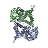


 PDBj
PDBj


 Assembly
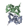


 Type: L-peptide linking / Mass: 147.129 Da / Num. of mol.: 2 / Source method: obtained synthetically / Formula: C5H9NO4
 Mass: 35.453 Da / Num. of mol.: 5 / Source method: obtained synthetically / Formula: Cl
 Mass: 22.990 Da / Num. of mol.: 2 / Source method: obtained synthetically / Formula: Na
 Mass: 60.095 Da / Num. of mol.: 2 / Source method: obtained synthetically / Formula: C3H8O
 Sample preparation
 / Beamline: 22-ID / Wavelength: 1 Å
 Processing