[English] 日本語
 Yorodumi
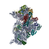
- PDB-5me1: Structure of the 30S Pre-Initiation Complex 2 (30S IC-2) Stalled ... -
+ Open data
- Basic information
| Entry | Database: PDB / ID: 5me1 | |||||||||||||||||||||||||||||||||||||||||||||||||||||||||||||||||||||||||||||||||||||||||||||||||||||||||||||||||||||||||||||||||||||||||||||||||||||||||
|---|---|---|---|---|---|---|---|---|---|---|---|---|---|---|---|---|---|---|---|---|---|---|---|---|---|---|---|---|---|---|---|---|---|---|---|---|---|---|---|---|---|---|---|---|---|---|---|---|---|---|---|---|---|---|---|---|---|---|---|---|---|---|---|---|---|---|---|---|---|---|---|---|---|---|---|---|---|---|---|---|---|---|---|---|---|---|---|---|---|---|---|---|---|---|---|---|---|---|---|---|---|---|---|---|---|---|---|---|---|---|---|---|---|---|---|---|---|---|---|---|---|---|---|---|---|---|---|---|---|---|---|---|---|---|---|---|---|---|---|---|---|---|---|---|---|---|---|---|---|---|---|---|---|---|
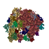
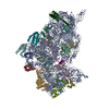
| Title | Structure of the 30S Pre-Initiation Complex 2 (30S IC-2) Stalled by GE81112 | |||||||||||||||||||||||||||||||||||||||||||||||||||||||||||||||||||||||||||||||||||||||||||||||||||||||||||||||||||||||||||||||||||||||||||||||||||||||||
 Components Components |
| |||||||||||||||||||||||||||||||||||||||||||||||||||||||||||||||||||||||||||||||||||||||||||||||||||||||||||||||||||||||||||||||||||||||||||||||||||||||||
 Keywords Keywords | RIBOSOME / Initiation of Translation | |||||||||||||||||||||||||||||||||||||||||||||||||||||||||||||||||||||||||||||||||||||||||||||||||||||||||||||||||||||||||||||||||||||||||||||||||||||||||
| Function / homology |  Function and homology information Function and homology informationribosome disassembly / guanosine tetraphosphate binding / : / transcription antitermination factor activity, RNA binding / ornithine decarboxylase inhibitor activity / ribosomal small subunit binding / misfolded RNA binding / Group I intron splicing / RNA folding / negative regulation of translational initiation ...ribosome disassembly / guanosine tetraphosphate binding / : / transcription antitermination factor activity, RNA binding / ornithine decarboxylase inhibitor activity / ribosomal small subunit binding / misfolded RNA binding / Group I intron splicing / RNA folding / negative regulation of translational initiation / regulation of mRNA stability / translation initiation factor activity / mRNA regulatory element binding translation repressor activity / response to cold / positive regulation of RNA splicing / regulation of DNA-templated transcription elongation / transcription elongation factor complex / transcription antitermination / translational initiation / DNA-templated transcription termination / maintenance of translational fidelity / mRNA 5'-UTR binding / regulation of translation / ribosome biogenesis / ribosome binding / ribosomal small subunit biogenesis / ribosomal small subunit assembly / small ribosomal subunit / small ribosomal subunit rRNA binding / cytosolic small ribosomal subunit / cytoplasmic translation / tRNA binding / negative regulation of translation / rRNA binding / structural constituent of ribosome / ribosome / translation / response to antibiotic / GTPase activity / mRNA binding / GTP binding / RNA binding / zinc ion binding / membrane / cytoplasm / cytosol Similarity search - Function | |||||||||||||||||||||||||||||||||||||||||||||||||||||||||||||||||||||||||||||||||||||||||||||||||||||||||||||||||||||||||||||||||||||||||||||||||||||||||
| Biological species |    Thermus thermophilus (bacteria) Thermus thermophilus (bacteria)  Geobacillus stearothermophilus (bacteria) Geobacillus stearothermophilus (bacteria) | |||||||||||||||||||||||||||||||||||||||||||||||||||||||||||||||||||||||||||||||||||||||||||||||||||||||||||||||||||||||||||||||||||||||||||||||||||||||||
| Method | ELECTRON MICROSCOPY / single particle reconstruction / cryo EM / Resolution: 13.5 Å | |||||||||||||||||||||||||||||||||||||||||||||||||||||||||||||||||||||||||||||||||||||||||||||||||||||||||||||||||||||||||||||||||||||||||||||||||||||||||
 Authors Authors | Lopez-Alonso, J.P. / Fabbretti, A. / Kaminishi, T. / Iturrioz, I. / Brandi, L. / Gil Carton, D. / Gualerzi, C. / Fucini, P. / Connell, S. | |||||||||||||||||||||||||||||||||||||||||||||||||||||||||||||||||||||||||||||||||||||||||||||||||||||||||||||||||||||||||||||||||||||||||||||||||||||||||
 Citation Citation |  Journal: Nucleic Acids Res / Year: 2017 Journal: Nucleic Acids Res / Year: 2017Title: Structure of a 30S pre-initiation complex stalled by GE81112 reveals structural parallels in bacterial and eukaryotic protein synthesis initiation pathways. Authors: Jorge P López-Alonso / Attilio Fabbretti / Tatsuya Kaminishi / Idoia Iturrioz / Letizia Brandi / David Gil-Carton / Claudio O Gualerzi / Paola Fucini / Sean R Connell /   Abstract: In bacteria, the start site and the reading frame of the messenger RNA are selected by the small ribosomal subunit (30S) when the start codon, typically an AUG, is decoded in the P-site by the ...In bacteria, the start site and the reading frame of the messenger RNA are selected by the small ribosomal subunit (30S) when the start codon, typically an AUG, is decoded in the P-site by the initiator tRNA in a process guided and controlled by three initiation factors. This process can be efficiently inhibited by GE81112, a natural tetrapeptide antibiotic that is highly specific toward bacteria. Here GE81112 was used to stabilize the 30S pre-initiation complex and obtain its structure by cryo-electron microscopy. The results obtained reveal the occurrence of changes in both the ribosome conformation and initiator tRNA position that may play a critical role in controlling translational fidelity. Furthermore, the structure highlights similarities with the early steps of initiation in eukaryotes suggesting that shared structural features guide initiation in all kingdoms of life. | |||||||||||||||||||||||||||||||||||||||||||||||||||||||||||||||||||||||||||||||||||||||||||||||||||||||||||||||||||||||||||||||||||||||||||||||||||||||||
| History |
|
- Structure visualization
| Movie |
 Movie viewer Movie viewer |
|---|---|
| Structure viewer | Molecule:  Molmil Molmil Jmol/JSmol Jmol/JSmol |
- Downloads & links
- Download
| PDBx/mmCIF format |  5me1.cif.gz 5me1.cif.gz | 1.5 MB | Display |  PDBx/mmCIF format PDBx/mmCIF format |
|---|---|---|---|---|
| PDB format |  pdb5me1.ent.gz pdb5me1.ent.gz | 1.1 MB | Display |  PDB format PDB format |
| PDBx/mmJSON format |  5me1.json.gz 5me1.json.gz | Tree view |  PDBx/mmJSON format PDBx/mmJSON format | |
| Others |  Other downloads Other downloads |
-Validation report
| Arichive directory |  https://data.pdbj.org/pub/pdb/validation_reports/me/5me1 https://data.pdbj.org/pub/pdb/validation_reports/me/5me1 ftp://data.pdbj.org/pub/pdb/validation_reports/me/5me1 ftp://data.pdbj.org/pub/pdb/validation_reports/me/5me1 | HTTPS FTP |
|---|
-Related structure data
| Related structure data |  3495MC  3494C  5me0C M: map data used to model this data C: citing same article ( |
|---|---|
| Similar structure data |
- Links
Links
- Assembly
| Deposited unit | 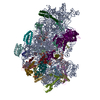
|
|---|---|
| 1 |
|
- Components
-RNA chain , 2 types, 2 molecules AX
| #1: RNA chain | Mass: 497404.969 Da / Num. of mol.: 1 / Source method: isolated from a natural source / Details: Taken from PDB entry 4YBB / Source: (natural)  |
|---|---|
| #26: RNA chain | Mass: 24818.893 Da / Num. of mol.: 1 / Source method: isolated from a natural source / Details: Taken from PDB entry 3JCN / Source: (natural)  |
-30S ribosomal protein ... , 20 types, 20 molecules BCDEFGHIJKLMNOPQRSTU
| #2: Protein | Mass: 26781.670 Da / Num. of mol.: 1 / Source method: isolated from a natural source / Details: Taken from PDB entry 4YBB / Source: (natural)  |
|---|---|
| #3: Protein | Mass: 26031.316 Da / Num. of mol.: 1 / Source method: isolated from a natural source / Details: Taken from PDB entry 4YBB / Source: (natural)  |
| #4: Protein | Mass: 23514.199 Da / Num. of mol.: 1 / Source method: isolated from a natural source / Details: Taken from PDB entry 4YBB / Source: (natural)  |
| #5: Protein | Mass: 17629.398 Da / Num. of mol.: 1 / Source method: isolated from a natural source / Details: Taken from PDB entry 4YBB / Source: (natural)  |
| #6: Protein | Mass: 15211.058 Da / Num. of mol.: 1 / Source method: isolated from a natural source / Source: (natural)  |
| #7: Protein | Mass: 17637.445 Da / Num. of mol.: 1 / Source method: isolated from a natural source / Details: Taken from PDB entry 4YBB / Source: (natural)  |
| #8: Protein | Mass: 14146.557 Da / Num. of mol.: 1 / Source method: isolated from a natural source / Details: Taken from PDB entry 4YBB / Source: (natural)  |
| #9: Protein | Mass: 14886.270 Da / Num. of mol.: 1 / Source method: isolated from a natural source / Details: Taken from PDB entry 4YBB / Source: (natural)  |
| #10: Protein | Mass: 11755.597 Da / Num. of mol.: 1 / Source method: isolated from a natural source / Details: Taken from PDB entry 4YBB / Source: (natural)  |
| #11: Protein | Mass: 13870.975 Da / Num. of mol.: 1 / Source method: isolated from a natural source / Details: Taken from PDB entry 4YBB / Source: (natural)  |
| #12: Protein | Mass: 13683.053 Da / Num. of mol.: 1 / Source method: isolated from a natural source / Details: Taken from PDB entry 4YBB / Source: (natural)  |
| #13: Protein | Mass: 13128.467 Da / Num. of mol.: 1 Source method: isolated from a genetically manipulated source Details: Taken from PDB entry 4YBB / Source: (gene. exp.)   |
| #14: Protein | Mass: 11606.560 Da / Num. of mol.: 1 / Source method: isolated from a natural source / Details: Taken from PDB entry 4YBB / Source: (natural)  |
| #15: Protein | Mass: 10290.816 Da / Num. of mol.: 1 / Source method: isolated from a natural source / Details: Taken from PDB entry 4YBB / Source: (natural)  |
| #16: Protein | Mass: 11464.126 Da / Num. of mol.: 1 / Source method: isolated from a natural source / Details: Taken from PDB entry 4YBB / Source: (natural)  |
| #17: Protein | Mass: 9724.491 Da / Num. of mol.: 1 / Source method: isolated from a natural source / Details: Taken from PDB entry 4YBB / Source: (natural)  |
| #18: Protein | Mass: 9005.472 Da / Num. of mol.: 1 / Source method: isolated from a natural source / Details: Taken from PDB entry 4YBB / Source: (natural)  |
| #19: Protein | Mass: 10455.355 Da / Num. of mol.: 1 / Source method: isolated from a natural source / Details: Taken from PDB entry 4YBB / Source: (natural)  |
| #20: Protein | Mass: 9708.464 Da / Num. of mol.: 1 / Source method: isolated from a natural source / Details: Taken from PDB entry 4YBB / Source: (natural)  |
| #21: Protein | Mass: 8524.039 Da / Num. of mol.: 1 / Source method: isolated from a natural source / Details: Taken from PDB entry 4YBB / Source: (natural)  |
-Translation initiation factor IF- ... , 4 types, 4 molecules VWYZ
| #22: Protein | Mass: 8247.664 Da / Num. of mol.: 1 Source method: isolated from a genetically manipulated source Details: Taken from PDB entry 1HR0 Source: (gene. exp.)   Thermus thermophilus (strain HB8 / ATCC 27634 / DSM 579) (bacteria) Thermus thermophilus (strain HB8 / ATCC 27634 / DSM 579) (bacteria)Strain: HB8 / ATCC 27634 / DSM 579 / Gene: infA, TTHA1669 / Production host:  |
|---|---|
| #23: Protein | Mass: 97498.164 Da / Num. of mol.: 1 Source method: isolated from a genetically manipulated source Details: Taken from PDB entry 3JCN / Source: (gene. exp.)   |
| #24: Protein | Mass: 19717.984 Da / Num. of mol.: 1 Source method: isolated from a genetically manipulated source Details: Taken from PDB entry 1TIF Source: (gene. exp.)   Geobacillus stearothermophilus (bacteria) Geobacillus stearothermophilus (bacteria)Gene: infC / Production host:  |
| #25: Protein | Mass: 16667.477 Da / Num. of mol.: 1 Source method: isolated from a genetically manipulated source Details: Taken from PDB entry 2IFE / Source: (gene. exp.)   |
-Non-polymers , 1 types, 1 molecules 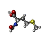
| #27: Chemical | ChemComp-FME / |
|---|
-Details
| Has protein modification | Y |
|---|
-Experimental details
-Experiment
| Experiment | Method: ELECTRON MICROSCOPY |
|---|---|
| EM experiment | Aggregation state: PARTICLE / 3D reconstruction method: single particle reconstruction |
- Sample preparation
| Component | Name: 30S Pre-Initiation Complex IC-2 Stalled by GE81112 / Type: RIBOSOME / Entity ID: #1-#26 / Source: MULTIPLE SOURCES | ||||||||||||||||||||
|---|---|---|---|---|---|---|---|---|---|---|---|---|---|---|---|---|---|---|---|---|---|
| Molecular weight | Experimental value: NO | ||||||||||||||||||||
| Source (natural) | Organism:  | ||||||||||||||||||||
| Buffer solution | pH: 7.7 | ||||||||||||||||||||
| Buffer component |
| ||||||||||||||||||||
| Specimen | Embedding applied: NO / Shadowing applied: NO / Staining applied: NO / Vitrification applied: YES | ||||||||||||||||||||
| Specimen support | Grid material: COPPER / Grid mesh size: 200 divisions/in. / Grid type: Quantifoil R2/1 | ||||||||||||||||||||
| Vitrification | Instrument: FEI VITROBOT MARK II / Cryogen name: ETHANE / Humidity: 95 % / Chamber temperature: 278 K |
- Electron microscopy imaging
| Microscopy | Model: JEOL 2200FSC |
|---|---|
| Electron gun | Electron source:  FIELD EMISSION GUN / Accelerating voltage: 200 kV / Illumination mode: FLOOD BEAM FIELD EMISSION GUN / Accelerating voltage: 200 kV / Illumination mode: FLOOD BEAM |
| Electron lens | Mode: BRIGHT FIELD / Nominal magnification: 50000 X / Calibrated magnification: 74183 X / Cs: 2 mm |
| Specimen holder | Cryogen: NITROGEN / Specimen holder model: GATAN LIQUID NITROGEN |
| Image recording | Average exposure time: 0.3 sec. / Electron dose: 17 e/Å2 / Film or detector model: GATAN ULTRASCAN 4000 (4k x 4k) / Num. of grids imaged: 1 / Num. of real images: 261 |
| EM imaging optics | Energyfilter name: In-column Omega Filter |
| Image scans | Width: 4096 / Height: 4096 |
- Processing
| EM software |
| ||||||||||||||||||||||||||||||||||||||||||||||||||
|---|---|---|---|---|---|---|---|---|---|---|---|---|---|---|---|---|---|---|---|---|---|---|---|---|---|---|---|---|---|---|---|---|---|---|---|---|---|---|---|---|---|---|---|---|---|---|---|---|---|---|---|
| CTF correction | Type: PHASE FLIPPING AND AMPLITUDE CORRECTION | ||||||||||||||||||||||||||||||||||||||||||||||||||
| Particle selection | Num. of particles selected: 174142 | ||||||||||||||||||||||||||||||||||||||||||||||||||
| Symmetry | Point symmetry: C1 (asymmetric) | ||||||||||||||||||||||||||||||||||||||||||||||||||
| 3D reconstruction | Resolution: 13.5 Å / Resolution method: FSC 0.143 CUT-OFF / Num. of particles: 26776 / Num. of class averages: 1 / Symmetry type: POINT | ||||||||||||||||||||||||||||||||||||||||||||||||||
| Atomic model building | Protocol: RIGID BODY FIT Details: The fitting of the model to the EM volume was performed through rigid body fitting. The head and the body of the 30S were treated as independent bodies. Due to the low resolution of the ...Details: The fitting of the model to the EM volume was performed through rigid body fitting. The head and the body of the 30S were treated as independent bodies. Due to the low resolution of the volume, the conformation of the linking bases was not minimized. In addition, some of the clashes of the model are produced by flexible loops or protein side chains that were not refined. | ||||||||||||||||||||||||||||||||||||||||||||||||||
| Atomic model building | 3D fitting-ID: 1 / Source name: PDB / Type: experimental model
|
 Movie
Movie Controller



 PDBj
PDBj


