 Movie
Movie Controller

[English] 日本語
 Yorodumi
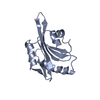
- PDB-5lsl: Crystal structure of yeast Hsh49p in complex with Cus1p binding d... -
+ Open data

- Basic information
| Entry | Database: PDB / ID: 5lsl | ||||||
|---|---|---|---|---|---|---|---|
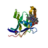
| Title | Crystal structure of yeast Hsh49p in complex with Cus1p binding domain. | ||||||
 Components Components |
| ||||||
 Keywords Keywords | RNA binding domain / Splicing / U2 snRNP / SF3b complex | ||||||
| Function / homology |  Function and homology information Function and homology informationU2-type prespliceosome assembly / U2-type spliceosomal complex / U2 snRNP / U2-type prespliceosome / precatalytic spliceosome / spliceosomal complex assembly / Prp19 complex / catalytic step 2 spliceosome / spliceosomal complex / mRNA splicing, via spliceosome ...U2-type prespliceosome assembly / U2-type spliceosomal complex / U2 snRNP / U2-type prespliceosome / precatalytic spliceosome / spliceosomal complex assembly / Prp19 complex / catalytic step 2 spliceosome / spliceosomal complex / mRNA splicing, via spliceosome / RNA binding / nucleus Similarity search - Function | ||||||
| Biological species |  | ||||||
| Method |  X-RAY DIFFRACTION / SYNCHROTRON / MOLECULAR REPLACEMENT / molecular replacement / Resolution: 1.65 Å X-RAY DIFFRACTION / SYNCHROTRON / MOLECULAR REPLACEMENT / molecular replacement / Resolution: 1.65 Å | ||||||
 Authors Authors | van Roon, A.M. / Obayashi, E. / Sposito, B. / Oubridge, C. / Nagai, K. | ||||||
 Citation Citation | Journal: RNA / Year: 2017 Title: Crystal structure of U2 snRNP SF3b components: Hsh49p in complex with Cus1p-binding domain. Authors: van Roon, A.M. / Oubridge, C. / Obayashi, E. / Sposito, B. / Newman, A.J. / Seraphin, B. / Nagai, K. | ||||||
| History |
|
- Structure visualization
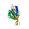
| Structure viewer | Molecule: MolmilJmol/JSmol |
|---|
- Downloads & links
-Download
| PDBx/mmCIF format | 5lsl.cif.gz | 242 KB | Display | PDBx/mmCIF format |
|---|---|---|---|---|
| PDB format | pdb5lsl.ent.gz | 196.6 KB | Display | PDB format |
| PDBx/mmJSON format | 5lsl.json.gz | Tree view | PDBx/mmJSON format | |
| Others |  Other downloads Other downloads |
-Validation report
| Arichive directory | https://data.pdbj.org/pub/pdb/validation_reports/ls/5lslftp://data.pdbj.org/pub/pdb/validation_reports/ls/5lsl | HTTPS FTP |
|---|
-Related structure data
| Related structure data |  5lsbC  3mdfS C: citing same article ( S: Starting model for refinement |
|---|---|
| Similar structure data |


-Links
 PDBj
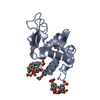
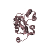
PDBj- Assembly
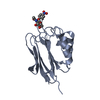
| Deposited unit | 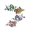
| ||||||||
|---|---|---|---|---|---|---|---|---|---|
| 1 | 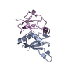
| ||||||||
| 2 | 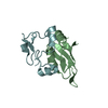
| ||||||||
| 3 | 
| ||||||||
| 4 | 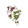
| ||||||||
| Unit cell |
|
-Components
| #1: Protein | Mass: 11612.056 Da / Num. of mol.: 4 Source method: isolated from a genetically manipulated source Source: (gene. exp.) Gene: HSH49, YOR319W, O6142 / Plasmid: pRK-172 / Details (production host): His-tag with a TEV cleavage site / Production host:  #2: Protein | Mass: 9388.993 Da / Num. of mol.: 4 Source method: isolated from a genetically manipulated source Source: (gene. exp.) Gene: CUS1, YMR240C, YM9408.02C / Plasmid: pRK-172 Details (production host): His-tagged protein with TEV cleavage site Production host: #3: Water | ChemComp-HOH / |  Mass: 18.015 Da / Num. of mol.: 402 / Source method: isolated from a natural source / Formula: H2O Mass: 18.015 Da / Num. of mol.: 402 / Source method: isolated from a natural source / Formula: H2O |
|---|
-Experimental details
-Experiment
| Experiment | Method: X-RAY DIFFRACTION / Number of used crystals: 1 |
|---|
- Sample preparation
| Crystal | Density Matthews: 2.23 Å3/Da / Density % sol: 44.75 % / Description: thin plate with dimensions 0.25x0.1 mm |
|---|---|
| Crystal grow | Temperature: 293 K / Method: vapor diffusion, sitting drop / pH: 8.5 / Details: 18% PEG4000, 0.1M Tris HCl pH 8.5, 45 mM LiSO4 / PH range: 8.0-8.5 |
-Data collection
| Diffraction | Mean temperature: 100 K | ||||||||||||||||||
|---|---|---|---|---|---|---|---|---|---|---|---|---|---|---|---|---|---|---|---|
| Diffraction source | Source: SYNCHROTRON / Site: Diamond  / Beamline: I02 / Wavelength: 0.98 Å / Beamline: I02 / Wavelength: 0.98 Å | ||||||||||||||||||
| Detector | Type: DECTRIS PILATUS3 S 6M / Detector: PIXEL / Date: Feb 22, 2013 | ||||||||||||||||||
| Radiation | Protocol: SINGLE WAVELENGTH / Monochromatic (M) / Laue (L): M / Scattering type: x-ray | ||||||||||||||||||
| Radiation wavelength | Wavelength: 0.98 Å / Relative weight: 1 | ||||||||||||||||||
| Reflection | Resolution: 1.65→44.47 Å / Num. obs: 84322 / % possible obs: 96.7 % / Redundancy: 2.3 % / CC1/2: 0.988 / Rmerge(I) obs: 0.077 / Net I/σ(I): 13.3 | ||||||||||||||||||
| Reflection shell |
|
-Phasing
| Phasing | Method: molecular replacement |
|---|
- Processing
| Software |
| |||||||||||||||||||||||||||||||||||||||||||||||||||||||||||||||||||||||||||||||||||||||||||||||||||||||||||||||||||||||||||||||||||||||||||||||||||||||||||||||||||||||||||||||||||||||||||||||||||||||||||||||||||||||||||||||||
|---|---|---|---|---|---|---|---|---|---|---|---|---|---|---|---|---|---|---|---|---|---|---|---|---|---|---|---|---|---|---|---|---|---|---|---|---|---|---|---|---|---|---|---|---|---|---|---|---|---|---|---|---|---|---|---|---|---|---|---|---|---|---|---|---|---|---|---|---|---|---|---|---|---|---|---|---|---|---|---|---|---|---|---|---|---|---|---|---|---|---|---|---|---|---|---|---|---|---|---|---|---|---|---|---|---|---|---|---|---|---|---|---|---|---|---|---|---|---|---|---|---|---|---|---|---|---|---|---|---|---|---|---|---|---|---|---|---|---|---|---|---|---|---|---|---|---|---|---|---|---|---|---|---|---|---|---|---|---|---|---|---|---|---|---|---|---|---|---|---|---|---|---|---|---|---|---|---|---|---|---|---|---|---|---|---|---|---|---|---|---|---|---|---|---|---|---|---|---|---|---|---|---|---|---|---|---|---|---|---|---|---|---|---|---|---|---|---|---|---|---|---|---|---|---|---|---|
| Refinement | Method to determine structure: MOLECULAR REPLACEMENT Starting model: 3MDF Resolution: 1.65→44.47 Å / Cor.coef. Fo:Fc: 0.944 / Cor.coef. Fo:Fc free: 0.93 / SU B: 3.978 / SU ML: 0.068 / SU R Cruickshank DPI: 0.0955 / Cross valid method: FREE R-VALUE / σ(F): 0 / ESU R: 0.095 / ESU R Free: 0.092 / Details: HYDROGENS HAVE BEEN ADDED IN THE RIDING POSITIONS
| |||||||||||||||||||||||||||||||||||||||||||||||||||||||||||||||||||||||||||||||||||||||||||||||||||||||||||||||||||||||||||||||||||||||||||||||||||||||||||||||||||||||||||||||||||||||||||||||||||||||||||||||||||||||||||||||||
| Solvent computation | Ion probe radii: 0.8 Å / Shrinkage radii: 0.8 Å / VDW probe radii: 1.1 Å | |||||||||||||||||||||||||||||||||||||||||||||||||||||||||||||||||||||||||||||||||||||||||||||||||||||||||||||||||||||||||||||||||||||||||||||||||||||||||||||||||||||||||||||||||||||||||||||||||||||||||||||||||||||||||||||||||
| Displacement parameters | Biso max: 79.41 Å2 / Biso mean: 18.689 Å2 / Biso min: 6.32 Å2
| |||||||||||||||||||||||||||||||||||||||||||||||||||||||||||||||||||||||||||||||||||||||||||||||||||||||||||||||||||||||||||||||||||||||||||||||||||||||||||||||||||||||||||||||||||||||||||||||||||||||||||||||||||||||||||||||||
| Refinement step | Cycle: final / Resolution: 1.65→44.47 Å
| |||||||||||||||||||||||||||||||||||||||||||||||||||||||||||||||||||||||||||||||||||||||||||||||||||||||||||||||||||||||||||||||||||||||||||||||||||||||||||||||||||||||||||||||||||||||||||||||||||||||||||||||||||||||||||||||||
| Refine LS restraints |
| |||||||||||||||||||||||||||||||||||||||||||||||||||||||||||||||||||||||||||||||||||||||||||||||||||||||||||||||||||||||||||||||||||||||||||||||||||||||||||||||||||||||||||||||||||||||||||||||||||||||||||||||||||||||||||||||||
| LS refinement shell | Resolution: 1.65→1.693 Å / Total num. of bins used: 20
| |||||||||||||||||||||||||||||||||||||||||||||||||||||||||||||||||||||||||||||||||||||||||||||||||||||||||||||||||||||||||||||||||||||||||||||||||||||||||||||||||||||||||||||||||||||||||||||||||||||||||||||||||||||||||||||||||
| Refinement TLS params. | Method: refined / Refine-ID: X-RAY DIFFRACTION
| |||||||||||||||||||||||||||||||||||||||||||||||||||||||||||||||||||||||||||||||||||||||||||||||||||||||||||||||||||||||||||||||||||||||||||||||||||||||||||||||||||||||||||||||||||||||||||||||||||||||||||||||||||||||||||||||||
| Refinement TLS group |
|
