 Movie
Movie Controller

[English] 日本語
 Yorodumi
- PDB-5lsb: Crystal structure of yeast Hsh49p in complex with Cus1p binding d... -
+ Open data

- Basic information
| Entry | Database: PDB / ID: 5lsb | ||||||
|---|---|---|---|---|---|---|---|
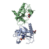
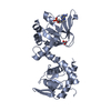
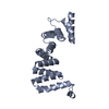
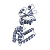
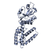
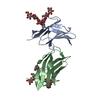
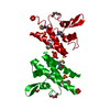
| Title | Crystal structure of yeast Hsh49p in complex with Cus1p binding domain. | ||||||
 Components Components |
| ||||||
 Keywords Keywords | RNA binding domain / Splicing / U2 snRNP / SF3b complex | ||||||
| Function / homology |  Function and homology information Function and homology informationU2-type prespliceosome assembly / U2-type spliceosomal complex / U2 snRNP / U2-type prespliceosome / precatalytic spliceosome / spliceosomal complex assembly / Prp19 complex / catalytic step 2 spliceosome / spliceosomal complex / mRNA splicing, via spliceosome ...U2-type prespliceosome assembly / U2-type spliceosomal complex / U2 snRNP / U2-type prespliceosome / precatalytic spliceosome / spliceosomal complex assembly / Prp19 complex / catalytic step 2 spliceosome / spliceosomal complex / mRNA splicing, via spliceosome / RNA binding / nucleus Similarity search - Function | ||||||
| Biological species |  | ||||||
| Method |  X-RAY DIFFRACTION / SYNCHROTRON / MOLECULAR REPLACEMENT / molecular replacement / Resolution: 2.7 Å X-RAY DIFFRACTION / SYNCHROTRON / MOLECULAR REPLACEMENT / molecular replacement / Resolution: 2.7 Å | ||||||
 Authors Authors | van Roon, A.M. / Obayashi, E. / Sposito, B. / Oubridge, C. / Nagai, K. | ||||||
 Citation Citation | Journal: RNA / Year: 2017 Title: Crystal structure of U2 snRNP SF3b components: Hsh49p in complex with Cus1p-binding domain. Authors: van Roon, A.M. / Oubridge, C. / Obayashi, E. / Sposito, B. / Newman, A.J. / Seraphin, B. / Nagai, K. | ||||||
| History |
|
- Structure visualization
| Structure viewer | Molecule: MolmilJmol/JSmol |
|---|
- Downloads & links
-Download
| PDBx/mmCIF format | 5lsb.cif.gz | 293.6 KB | Display | PDBx/mmCIF format |
|---|---|---|---|---|
| PDB format | pdb5lsb.ent.gz | 240.7 KB | Display | PDB format |
| PDBx/mmJSON format | 5lsb.json.gz | Tree view | PDBx/mmJSON format | |
| Others |  Other downloads Other downloads |
-Validation report
| Arichive directory | https://data.pdbj.org/pub/pdb/validation_reports/ls/5lsbftp://data.pdbj.org/pub/pdb/validation_reports/ls/5lsb | HTTPS FTP |
|---|
-Related structure data
| Related structure data | 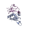 5lslSC 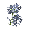 1cvjS S: Starting model for refinement C: citing same article ( |
|---|---|
| Similar structure data |


-Links
 PDBj
PDBj- Assembly
| Deposited unit | 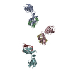
| ||||||||
|---|---|---|---|---|---|---|---|---|---|
| 1 | 
| ||||||||
| 2 | 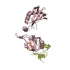
| ||||||||
| 3 | 
| ||||||||
| Unit cell |
|
-Components
| #1: Protein | Mass: 24604.141 Da / Num. of mol.: 3 Source method: isolated from a genetically manipulated source Details: GGS sequence at the N-terminus is non-natural and left over from TEV-protease cleavage site Source: (gene. exp.) Strain: ATCC 204508 / S288c / Gene: HSH49, YOR319W, O6142 / Production host:  #2: Protein | Mass: 9388.993 Da / Num. of mol.: 3 Source method: isolated from a genetically manipulated source Details: Hsh49p binding domain of Cus1p between residues 290-368 Source: (gene. exp.) Strain: ATCC 204508 / S288c / Gene: CUS1, YMR240C, YM9408.02C / Production host: #3: Water | ChemComp-HOH / |  Mass: 18.015 Da / Num. of mol.: 6 / Source method: isolated from a natural source / Formula: H2O Mass: 18.015 Da / Num. of mol.: 6 / Source method: isolated from a natural source / Formula: H2O |
|---|
-Experimental details
-Experiment
| Experiment | Method: X-RAY DIFFRACTION / Number of used crystals: 1 |
|---|
- Sample preparation
| Crystal | Density Matthews: 2.31 Å3/Da / Density % sol: 46.66 % / Description: Thin needles |
|---|---|
| Crystal grow | Temperature: 293 K / Method: vapor diffusion, sitting drop Details: Equal volumes of 10-15 mg/ml protein solution were mixed with an equal volume of reservoir solution containing 2.45-2.65 M NaCl in 0.1M sodium acetate pH 4.8-4.9 PH range: 4.8-4.9 |
-Data collection
| Diffraction | Mean temperature: 100 K | ||||||||||||||||||
|---|---|---|---|---|---|---|---|---|---|---|---|---|---|---|---|---|---|---|---|
| Diffraction source | Source: SYNCHROTRON / Site: ESRF  / Beamline: ID14-3 / Wavelength: 0.931 Å / Beamline: ID14-3 / Wavelength: 0.931 Å | ||||||||||||||||||
| Detector | Type: ADSC QUANTUM 4 / Detector: CCD / Date: Jul 6, 2007 | ||||||||||||||||||
| Radiation | Protocol: SINGLE WAVELENGTH / Monochromatic (M) / Laue (L): M / Scattering type: x-ray | ||||||||||||||||||
| Radiation wavelength | Wavelength: 0.931 Å / Relative weight: 1 | ||||||||||||||||||
| Reflection twin |
| ||||||||||||||||||
| Reflection | Resolution: 2.7→35.52 Å / Num. obs: 25001 / % possible obs: 99.9 % / Redundancy: 4.2 % / CC1/2: 0.984 / Rmerge(I) obs: 0.149 / Rpim(I) all: 0.083 / Rrim(I) all: 0.171 / Net I/σ(I): 13 / Num. measured all: 104708 / Scaling rejects: 11 | ||||||||||||||||||
| Reflection shell |
|
-Phasing
| Phasing | Method: molecular replacement | |||||||||
|---|---|---|---|---|---|---|---|---|---|---|
| Phasing MR | Model details: Phaser MODE: MR_AUTO
|
- Processing
| Software |
| |||||||||||||||||||||||||||||||||||||||||||||||||||||||||||||||||||||||||||||||||||||||||||||||||||||||||||||||||||||||||||||||||||||||||||||||||||||||||||||||||||||||||||||||
|---|---|---|---|---|---|---|---|---|---|---|---|---|---|---|---|---|---|---|---|---|---|---|---|---|---|---|---|---|---|---|---|---|---|---|---|---|---|---|---|---|---|---|---|---|---|---|---|---|---|---|---|---|---|---|---|---|---|---|---|---|---|---|---|---|---|---|---|---|---|---|---|---|---|---|---|---|---|---|---|---|---|---|---|---|---|---|---|---|---|---|---|---|---|---|---|---|---|---|---|---|---|---|---|---|---|---|---|---|---|---|---|---|---|---|---|---|---|---|---|---|---|---|---|---|---|---|---|---|---|---|---|---|---|---|---|---|---|---|---|---|---|---|---|---|---|---|---|---|---|---|---|---|---|---|---|---|---|---|---|---|---|---|---|---|---|---|---|---|---|---|---|---|---|---|---|---|
| Refinement | Method to determine structure: MOLECULAR REPLACEMENT Starting model: 5LSL, 1CVJ Resolution: 2.7→35.52 Å / Cor.coef. Fo:Fc: 0.9 / Cor.coef. Fo:Fc free: 0.847 / SU B: 20.317 / SU ML: 0.204 / SU R Cruickshank DPI: 0.3657 / Cross valid method: FREE R-VALUE / σ(F): 0 / ESU R: 0.366 / ESU R Free: 0.067 / Details: HYDROGENS HAVE BEEN ADDED IN THE RIDING POSITIONS
| |||||||||||||||||||||||||||||||||||||||||||||||||||||||||||||||||||||||||||||||||||||||||||||||||||||||||||||||||||||||||||||||||||||||||||||||||||||||||||||||||||||||||||||||
| Solvent computation | Ion probe radii: 0.8 Å / Shrinkage radii: 0.8 Å / VDW probe radii: 1.2 Å | |||||||||||||||||||||||||||||||||||||||||||||||||||||||||||||||||||||||||||||||||||||||||||||||||||||||||||||||||||||||||||||||||||||||||||||||||||||||||||||||||||||||||||||||
| Displacement parameters | Biso max: 86.82 Å2 / Biso mean: 30.827 Å2 / Biso min: 9.01 Å2
| |||||||||||||||||||||||||||||||||||||||||||||||||||||||||||||||||||||||||||||||||||||||||||||||||||||||||||||||||||||||||||||||||||||||||||||||||||||||||||||||||||||||||||||||
| Refinement step | Cycle: final / Resolution: 2.7→35.52 Å
| |||||||||||||||||||||||||||||||||||||||||||||||||||||||||||||||||||||||||||||||||||||||||||||||||||||||||||||||||||||||||||||||||||||||||||||||||||||||||||||||||||||||||||||||
| Refine LS restraints |
| |||||||||||||||||||||||||||||||||||||||||||||||||||||||||||||||||||||||||||||||||||||||||||||||||||||||||||||||||||||||||||||||||||||||||||||||||||||||||||||||||||||||||||||||
| LS refinement shell | Resolution: 2.7→2.77 Å / Total num. of bins used: 20
| |||||||||||||||||||||||||||||||||||||||||||||||||||||||||||||||||||||||||||||||||||||||||||||||||||||||||||||||||||||||||||||||||||||||||||||||||||||||||||||||||||||||||||||||
| Refinement TLS params. | Method: refined / Refine-ID: X-RAY DIFFRACTION
| |||||||||||||||||||||||||||||||||||||||||||||||||||||||||||||||||||||||||||||||||||||||||||||||||||||||||||||||||||||||||||||||||||||||||||||||||||||||||||||||||||||||||||||||
| Refinement TLS group |
|
