 Movie
Movie Controller


+ Open data

- Basic information
| Entry | Database: PDB / ID: 5ln1 | ||||||
|---|---|---|---|---|---|---|---|
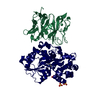
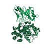
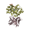
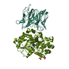
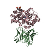
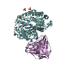
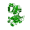
| Title | STRUCTURE OF UBIQUITYLATED-RPN10 FROM YEAST; | ||||||
 Components Components |
| ||||||
 Keywords Keywords | UBIQUITIN-BINDING PROTEIN / UBIQUITIN / VWA / UBIQUITYLATION / PROTEASOME | ||||||
| Function / homology |  Function and homology information Function and homology informationhypothalamus gonadotrophin-releasing hormone neuron development / female meiosis I / seminiferous tubule development / proteasome regulatory particle, base subcomplex / positive regulation of protein monoubiquitination / fat pad development / K48-linked polyubiquitin modification-dependent protein binding / mitochondrion transport along microtubule / female gonad development / male meiosis I ...hypothalamus gonadotrophin-releasing hormone neuron development / female meiosis I / seminiferous tubule development / proteasome regulatory particle, base subcomplex / positive regulation of protein monoubiquitination / fat pad development / K48-linked polyubiquitin modification-dependent protein binding / mitochondrion transport along microtubule / female gonad development / male meiosis I / positive regulation of intrinsic apoptotic signaling pathway by p53 class mediator / polyubiquitin modification-dependent protein binding / energy homeostasis / proteasome assembly / neuron projection morphogenesis / Maturation of protein E / Maturation of protein E / regulation of neuron apoptotic process / ER Quality Control Compartment (ERQC) / Myoclonic epilepsy of Lafora / FLT3 signaling by CBL mutants / IRAK2 mediated activation of TAK1 complex / Alpha-protein kinase 1 signaling pathway / Glycogen synthesis / IRAK1 recruits IKK complex / IRAK1 recruits IKK complex upon TLR7/8 or 9 stimulation / Prevention of phagosomal-lysosomal fusion / Endosomal Sorting Complex Required For Transport (ESCRT) / Membrane binding and targetting of GAG proteins / Regulation of TBK1, IKKε (IKBKE)-mediated activation of IRF3, IRF7 / Negative regulation of FLT3 / regulation of mitochondrial membrane potential / PTK6 Regulates RTKs and Their Effectors AKT1 and DOK1 / Regulation of TBK1, IKKε-mediated activation of IRF3, IRF7 upon TLR3 ligation / IRAK2 mediated activation of TAK1 complex upon TLR7/8 or 9 stimulation / Constitutive Signaling by NOTCH1 HD Domain Mutants / NOTCH2 Activation and Transmission of Signal to the Nucleus / TICAM1,TRAF6-dependent induction of TAK1 complex / TICAM1-dependent activation of IRF3/IRF7 / APC/C:Cdc20 mediated degradation of Cyclin B / Downregulation of ERBB4 signaling / APC-Cdc20 mediated degradation of Nek2A / Regulation of FZD by ubiquitination / proteasome complex / positive regulation of protein ubiquitination / p75NTR recruits signalling complexes / InlA-mediated entry of Listeria monocytogenes into host cells / regulation of proteasomal protein catabolic process / TRAF6 mediated IRF7 activation in TLR7/8 or 9 signaling / NF-kB is activated and signals survival / TRAF6-mediated induction of TAK1 complex within TLR4 complex / Regulation of pyruvate metabolism / Pexophagy / Downregulation of ERBB2:ERBB3 signaling / NRIF signals cell death from the nucleus / Regulation of PTEN localization / Regulation of innate immune responses to cytosolic DNA / VLDLR internalisation and degradation / Activated NOTCH1 Transmits Signal to the Nucleus / Synthesis of active ubiquitin: roles of E1 and E2 enzymes / Translesion synthesis by REV1 / TICAM1, RIP1-mediated IKK complex recruitment / Regulation of BACH1 activity / Translesion synthesis by POLK / JNK (c-Jun kinases) phosphorylation and activation mediated by activated human TAK1 / InlB-mediated entry of Listeria monocytogenes into host cell / MAP3K8 (TPL2)-dependent MAPK1/3 activation / Activation of IRF3, IRF7 mediated by TBK1, IKKε (IKBKE) / Translesion synthesis by POLI / Downregulation of TGF-beta receptor signaling / Josephin domain DUBs / Gap-filling DNA repair synthesis and ligation in GG-NER / IKK complex recruitment mediated by RIP1 / PINK1-PRKN Mediated Mitophagy / TGF-beta receptor signaling in EMT (epithelial to mesenchymal transition) / TNFR1-induced NF-kappa-B signaling pathway / Regulation of activated PAK-2p34 by proteasome mediated degradation / TCF dependent signaling in response to WNT / Regulation of NF-kappa B signaling / activated TAK1 mediates p38 MAPK activation / Autodegradation of Cdh1 by Cdh1:APC/C / APC/C:Cdc20 mediated degradation of Securin / NOTCH3 Activation and Transmission of Signal to the Nucleus / Regulation of signaling by CBL / Negative regulators of DDX58/IFIH1 signaling / N-glycan trimming in the ER and Calnexin/Calreticulin cycle / Asymmetric localization of PCP proteins / Fanconi Anemia Pathway / Negative regulation of FGFR3 signaling / Ubiquitin-dependent degradation of Cyclin D / Deactivation of the beta-catenin transactivating complex / Peroxisomal protein import / SCF-beta-TrCP mediated degradation of Emi1 / NIK-->noncanonical NF-kB signaling / Stabilization of p53 / AUF1 (hnRNP D0) binds and destabilizes mRNA / TNFR2 non-canonical NF-kB pathway / Negative regulation of FGFR2 signaling / Negative regulation of FGFR4 signaling / Downregulation of SMAD2/3:SMAD4 transcriptional activity Similarity search - Function | ||||||
| Biological species |   Homo sapiens (human) Homo sapiens (human) | ||||||
| Method |  X-RAY DIFFRACTION / SYNCHROTRON / MOLECULAR REPLACEMENT / Resolution: 3.14 Å X-RAY DIFFRACTION / SYNCHROTRON / MOLECULAR REPLACEMENT / Resolution: 3.14 Å | ||||||
 Authors Authors | Keren-Kaplan, T. / Attali, I. / Levin-Kravets, O. / Prag, G. | ||||||
| Funding support |  Israel, 1items Israel, 1items
| ||||||
 Citation Citation | Journal: Nat Commun / Year: 2016 Title: Structure of ubiquitylated-Rpn10 provides insight into its autoregulation mechanism. Authors: Keren-Kaplan, T. / Zeev Peters, L. / Levin-Kravets, O. / Attali, I. / Kleifeld, O. / Shohat, N. / Artzi, S. / Zucker, O. / Pilzer, I. / Reis, N. / Glickman, M.H. / Ben-Aroya, S. / Prag, G. #1: Journal: Acta Crystallogr. Sect. F Struct. Biol. Cryst. Commun. Year: 2012 Title: Purification and crystallization of mono-ubiquitylated ubiquitin receptor Rpn10. Authors: Keren-Kaplan, T. / Prag, G. | ||||||
| History |
|
- Structure visualization
| Structure viewer | Molecule: MolmilJmol/JSmol |
|---|
- Downloads & links
-Download
| PDBx/mmCIF format | 5ln1.cif.gz | 64.7 KB | Display | PDBx/mmCIF format |
|---|---|---|---|---|
| PDB format | pdb5ln1.ent.gz | 46.5 KB | Display | PDB format |
| PDBx/mmJSON format | 5ln1.json.gz | Tree view | PDBx/mmJSON format | |
| Others |  Other downloads Other downloads |
-Validation report
| Arichive directory | https://data.pdbj.org/pub/pdb/validation_reports/ln/5ln1ftp://data.pdbj.org/pub/pdb/validation_reports/ln/5ln1 | HTTPS FTP |
|---|
-Related structure data


-Links
 PDBj
PDBj


- Assembly
| Deposited unit | 
| ||||||||
|---|---|---|---|---|---|---|---|---|---|
| 1 | 
| ||||||||
| Unit cell |
|
-Components
| #1: Protein | Mass: 21624.496 Da / Num. of mol.: 1 / Fragment: UNP RESIDUES 1-191 Source method: isolated from a genetically manipulated source Source: (gene. exp.) Strain: ATCC 204508 / S288c / Gene: RPN10, MCB1, SUN1, YHR200W / Production host:  |
|---|---|
| #2: Protein | Mass: 8980.285 Da / Num. of mol.: 1 Source method: isolated from a genetically manipulated source Source: (gene. exp.) Homo sapiens (human) / Gene: UBB / Production host: |
-Experimental details
-Experiment
| Experiment | Method: X-RAY DIFFRACTION / Number of used crystals: 1 |
|---|
- Sample preparation
| Crystal | Density Matthews: 2.69 Å3/Da / Density % sol: 54.3 % |
|---|---|
| Crystal grow | Temperature: 292 K / Method: evaporation, recrystallization / pH: 6.5 Details: 12% (W/V) PEG 20000, 0.1M MES PH 6.5, VAPOR DIFFUSION, SITTING DROP, TEMPERATURE 292K |
-Data collection
| Diffraction | Mean temperature: 100 K |
|---|---|
| Diffraction source | Source: SYNCHROTRON / Site: ESRF  / Beamline: ID14-4 / Wavelength: 0.9393 Å / Beamline: ID14-4 / Wavelength: 0.9393 Å |
| Detector | Type: ADSC QUANTUM 315r / Detector: CCD / Date: Nov 28, 2011 |
| Radiation | Protocol: SINGLE WAVELENGTH / Monochromatic (M) / Laue (L): M / Scattering type: x-ray |
| Radiation wavelength | Wavelength: 0.9393 Å / Relative weight: 1 |
| Reflection | Resolution: 3.14→61.8 Å / Num. obs: 5824 / % possible obs: 99.8 % / Observed criterion σ(I): 9.8 / Redundancy: 3.2 % / Rmerge(I) obs: 0.084 / Rsym value: 0.098 / Net I/σ(I): 10.2 |
| Reflection shell | Resolution: 3.14→3.31 Å / Redundancy: 3.4 % / Rmerge(I) obs: 0.225 / Mean I/σ(I) obs: 5.1 / % possible all: 99.9 |
- Processing
| Software |
| ||||||||||||||||||||||||
|---|---|---|---|---|---|---|---|---|---|---|---|---|---|---|---|---|---|---|---|---|---|---|---|---|---|
| Refinement | Method to determine structure: MOLECULAR REPLACEMENT Starting model: 2X5N, 1UBQ Resolution: 3.14→53.64 Å / SU ML: 0.24 / Cross valid method: FREE R-VALUE / σ(F): 1.35 / Phase error: 25.21
| ||||||||||||||||||||||||
| Solvent computation | Shrinkage radii: 0.9 Å / VDW probe radii: 1.11 Å | ||||||||||||||||||||||||
| Refinement step | Cycle: LAST / Resolution: 3.14→53.64 Å
| ||||||||||||||||||||||||
| Refine LS restraints |
| ||||||||||||||||||||||||
| LS refinement shell |
|
