 Movie
Movie Controller

[English] 日本語
 Yorodumi
- PDB-1ym5: Crystal structure of YHI9, the yeast member of the phenazine bios... -
+ Open data

- Basic information
| Entry | Database: PDB / ID: 1ym5 | ||||||
|---|---|---|---|---|---|---|---|
| Title | Crystal structure of YHI9, the yeast member of the phenazine biosynthesis PhzF enzyme superfamily. | ||||||
 Components Components | Hypothetical 32.6 kDa protein in DAP2-SLT2 intergenic region | ||||||
 Keywords Keywords | OXIDOREDUCTASE / PhzF enzyme superfamily / double hot-dog / Structural Genomics / Paris-Sud Yeast Structural Genomics / YSG | ||||||
| Function / homology |  Function and homology information Function and homology informationIsomerases; Racemases and epimerases / isomerase activity / endoplasmic reticulum unfolded protein response / chromatin Similarity search - Function | ||||||
| Biological species |  | ||||||
| Method |  X-RAY DIFFRACTION / SAD / Resolution: 2.05 Å X-RAY DIFFRACTION / SAD / Resolution: 2.05 Å | ||||||
 Authors Authors | Liger, D. / Quevillon-Cheruel, S. / Sorel, I. / Bremang, M. / Blondeau, K. / Aboulfath, I. / Janin, J. / Van Tilbeurgh, H. / Leulliot, N. / Paris-Sud Yeast Structural Genomics (YSG) | ||||||
 Citation Citation | Journal: Proteins / Year: 2005 Title: Crystal structure of YHI9, the yeast member of the phenazine biosynthesis PhzF enzyme superfamily Authors: Liger, D. / Quevillon-Cheruel, S. / Sorel, I. / Bremang, M. / Blondeau, K. / Aboulfath, I. / Janin, J. / van Tilbeurgh, H. / Leulliot, N. | ||||||
| History |
|
- Structure visualization
| Structure viewer | Molecule: MolmilJmol/JSmol |
|---|
- Downloads & links
-Download
| PDBx/mmCIF format | 1ym5.cif.gz | 72 KB | Display | PDBx/mmCIF format |
|---|---|---|---|---|
| PDB format | pdb1ym5.ent.gz | 53.4 KB | Display | PDB format |
| PDBx/mmJSON format | 1ym5.json.gz | Tree view | PDBx/mmJSON format | |
| Others |  Other downloads Other downloads |
-Validation report
| Arichive directory | https://data.pdbj.org/pub/pdb/validation_reports/ym/1ym5ftp://data.pdbj.org/pub/pdb/validation_reports/ym/1ym5 | HTTPS FTP |
|---|
-Related structure data
| Similar structure data | |
|---|---|
| Other databases |


-Links
 PDBj
PDBj- Assembly
| Deposited unit | 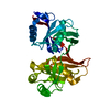
| ||||||||
|---|---|---|---|---|---|---|---|---|---|
| 1 | 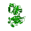
| ||||||||
| Unit cell |
|
-Components
| #1: Protein | Mass: 33427.238 Da / Num. of mol.: 1 Source method: isolated from a genetically manipulated source Source: (gene. exp.) Plasmid: pet9 / Production host:  |
|---|---|
| #2: Water | ChemComp-HOH /  Mass: 18.015 Da / Num. of mol.: 154 / Source method: isolated from a natural source / Formula: H2O Mass: 18.015 Da / Num. of mol.: 154 / Source method: isolated from a natural source / Formula: H2O |
-Experimental details
-Experiment
| Experiment | Method: X-RAY DIFFRACTION / Number of used crystals: 1 |
|---|
- Sample preparation
| Crystal | Density Matthews: 2 Å3/Da / Density % sol: 50 % |
|---|---|
| Crystal grow | Temperature: 293 K / Method: vapor diffusion, hanging drop / pH: 5 Details: 24% PEG 4000, 0.1M sodium acetate pH5, 0.2M ammonium acetate, 30% glycerol, 10mM DDT, VAPOR DIFFUSION, HANGING DROP, temperature 293K |
-Data collection
| Diffraction | Mean temperature: 100 K |
|---|---|
| Diffraction source | Source: ROTATING ANODE / Type: RIGAKU / Wavelength: 1.54 Å |
| Detector | Type: MARRESEARCH / Detector: IMAGE PLATE / Date: May 12, 2003 |
| Radiation | Monochromator: osmic / Protocol: SINGLE WAVELENGTH / Monochromatic (M) / Laue (L): M / Scattering type: x-ray |
| Radiation wavelength | Wavelength: 1.54 Å / Relative weight: 1 |
| Reflection | Resolution: 2.04→62.5 Å / Num. all: 21203 / Num. obs: 21044 / % possible obs: 98.4 % / Observed criterion σ(F): 2 / Observed criterion σ(I): 2 |
| Reflection shell | Resolution: 2.043→2.096 Å / % possible all: 92.69 |
- Processing
| Software |
| ||||||||||||||||||||||||||||||||||||||||||||||||||||||||||||||||||||||||||||||||||||||||||||||||||||||||||||||||||||||||||||||||||||||||||||||||||||||||||||||||||||||||||
|---|---|---|---|---|---|---|---|---|---|---|---|---|---|---|---|---|---|---|---|---|---|---|---|---|---|---|---|---|---|---|---|---|---|---|---|---|---|---|---|---|---|---|---|---|---|---|---|---|---|---|---|---|---|---|---|---|---|---|---|---|---|---|---|---|---|---|---|---|---|---|---|---|---|---|---|---|---|---|---|---|---|---|---|---|---|---|---|---|---|---|---|---|---|---|---|---|---|---|---|---|---|---|---|---|---|---|---|---|---|---|---|---|---|---|---|---|---|---|---|---|---|---|---|---|---|---|---|---|---|---|---|---|---|---|---|---|---|---|---|---|---|---|---|---|---|---|---|---|---|---|---|---|---|---|---|---|---|---|---|---|---|---|---|---|---|---|---|---|---|---|---|
| Refinement | Method to determine structure: SAD / Resolution: 2.05→40 Å / Cor.coef. Fo:Fc: 0.958 / Cor.coef. Fo:Fc free: 0.934 / SU B: 5.067 / SU ML: 0.137 / Cross valid method: THROUGHOUT / σ(F): 0.4 / ESU R: 0.193 / ESU R Free: 0.184 / Stereochemistry target values: MAXIMUM LIKELIHOOD / Details: HYDROGENS HAVE BEEN ADDED IN THE RIDING POSITIONS
| ||||||||||||||||||||||||||||||||||||||||||||||||||||||||||||||||||||||||||||||||||||||||||||||||||||||||||||||||||||||||||||||||||||||||||||||||||||||||||||||||||||||||||
| Solvent computation | Ion probe radii: 0.8 Å / Shrinkage radii: 0.8 Å / VDW probe radii: 1.2 Å / Solvent model: BABINET MODEL WITH MASK | ||||||||||||||||||||||||||||||||||||||||||||||||||||||||||||||||||||||||||||||||||||||||||||||||||||||||||||||||||||||||||||||||||||||||||||||||||||||||||||||||||||||||||
| Displacement parameters | Biso mean: 33.236 Å2
| ||||||||||||||||||||||||||||||||||||||||||||||||||||||||||||||||||||||||||||||||||||||||||||||||||||||||||||||||||||||||||||||||||||||||||||||||||||||||||||||||||||||||||
| Refinement step | Cycle: LAST / Resolution: 2.05→40 Å
| ||||||||||||||||||||||||||||||||||||||||||||||||||||||||||||||||||||||||||||||||||||||||||||||||||||||||||||||||||||||||||||||||||||||||||||||||||||||||||||||||||||||||||
| Refine LS restraints |
| ||||||||||||||||||||||||||||||||||||||||||||||||||||||||||||||||||||||||||||||||||||||||||||||||||||||||||||||||||||||||||||||||||||||||||||||||||||||||||||||||||||||||||
| LS refinement shell | Resolution: 2.043→2.096 Å / Total num. of bins used: 20
|
