 Movie
Movie Controller

[English] 日本語
 Yorodumi
- PDB-5li1: Structure of a Par3-inhibitory peptide bound to PKCiota core kina... -
+ Open data

- Basic information
| Entry | Database: PDB / ID: 5li1 | ||||||
|---|---|---|---|---|---|---|---|
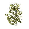
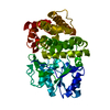
| Title | Structure of a Par3-inhibitory peptide bound to PKCiota core kinase domain | ||||||
 Components Components |
| ||||||
 Keywords Keywords | TRANSFERASE / aPKC / Polarity / Complex | ||||||
| Function / homology |  Function and homology information Function and homology informationdiacylglycerol-dependent, calcium-independent serine/threonine kinase activity / Golgi vesicle budding / PAR polarity complex / Tight junction interactions / protein kinase C / establishment of apical/basal cell polarity / diacylglycerol-dependent serine/threonine kinase activity / negative regulation of glial cell apoptotic process / eye photoreceptor cell development / Schmidt-Lanterman incisure ...diacylglycerol-dependent, calcium-independent serine/threonine kinase activity / Golgi vesicle budding / PAR polarity complex / Tight junction interactions / protein kinase C / establishment of apical/basal cell polarity / diacylglycerol-dependent serine/threonine kinase activity / negative regulation of glial cell apoptotic process / eye photoreceptor cell development / Schmidt-Lanterman incisure / establishment or maintenance of epithelial cell apical/basal polarity / membrane organization / cell-cell junction organization / cellular response to chemical stress / protein targeting to membrane / tight junction / positive regulation of Notch signaling pathway / establishment of cell polarity / cell leading edge / brush border / positive regulation of endothelial cell apoptotic process / positive regulation of glial cell proliferation / bicellular tight junction / regulation of postsynaptic membrane neurotransmitter receptor levels / intercellular bridge / vesicle-mediated transport / cytoskeleton organization / secretion / response to interleukin-1 / p75NTR recruits signalling complexes / positive regulation of D-glucose import across plasma membrane / : / actin filament organization / protein localization to plasma membrane / positive regulation of protein localization to plasma membrane / positive regulation of neuron projection development / phospholipid binding / Pre-NOTCH Transcription and Translation / Schaffer collateral - CA1 synapse / cellular response to insulin stimulus / KEAP1-NFE2L2 pathway / cell migration / microtubule cytoskeleton / protein phosphorylation / negative regulation of neuron apoptotic process / protein kinase activity / endosome / apical plasma membrane / intracellular signal transduction / cilium / Golgi membrane / protein serine kinase activity / protein serine/threonine kinase activity / negative regulation of apoptotic process / glutamatergic synapse / extracellular exosome / zinc ion binding / nucleoplasm / ATP binding / nucleus / plasma membrane / cytosol Similarity search - Function | ||||||
| Biological species |  Homo sapiens (human) Homo sapiens (human) | ||||||
| Method |  X-RAY DIFFRACTION / SYNCHROTRON / MOLECULAR REPLACEMENT / Resolution: 2 Å X-RAY DIFFRACTION / SYNCHROTRON / MOLECULAR REPLACEMENT / Resolution: 2 Å | ||||||
 Authors Authors | Soriano, E.V. / Purkiss, A.G. / McDonald, N.Q. | ||||||
 Citation Citation | Journal: Dev.Cell / Year: 2016 Title: aPKC Inhibition by Par3 CR3 Flanking Regions Controls Substrate Access and Underpins Apical-Junctional Polarization. Authors: Soriano, E.V. / Ivanova, M.E. / Fletcher, G. / Riou, P. / Knowles, P.P. / Barnouin, K. / Purkiss, A. / Kostelecky, B. / Saiu, P. / Linch, M. / Elbediwy, A. / Kjr, S. / O'Reilly, N. / ...Authors: Soriano, E.V. / Ivanova, M.E. / Fletcher, G. / Riou, P. / Knowles, P.P. / Barnouin, K. / Purkiss, A. / Kostelecky, B. / Saiu, P. / Linch, M. / Elbediwy, A. / Kjr, S. / O'Reilly, N. / Snijders, A.P. / Parker, P.J. / Thompson, B.J. / McDonald, N.Q. | ||||||
| History |
|
- Structure visualization
| Structure viewer | Molecule: MolmilJmol/JSmol |
|---|
- Downloads & links
-Download
| PDBx/mmCIF format | 5li1.cif.gz | 173.5 KB | Display | PDBx/mmCIF format |
|---|---|---|---|---|
| PDB format | pdb5li1.ent.gz | 133.5 KB | Display | PDB format |
| PDBx/mmJSON format | 5li1.json.gz | Tree view | PDBx/mmJSON format | |
| Others |  Other downloads Other downloads |
-Validation report
| Arichive directory | https://data.pdbj.org/pub/pdb/validation_reports/li/5li1ftp://data.pdbj.org/pub/pdb/validation_reports/li/5li1 | HTTPS FTP |
|---|
-Related structure data
| Related structure data |  5li9C  5lihC  3a8wS S: Starting model for refinement C: citing same article ( |
|---|---|
| Similar structure data |

-Links
 PDBj
PDBj


- Assembly
| Deposited unit | 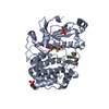
| ||||||||
|---|---|---|---|---|---|---|---|---|---|
| 1 |
| ||||||||
| Unit cell |
|
-Components
-Protein / Protein/peptide , 2 types, 2 molecules AB
| #1: Protein | Mass: 40914.934 Da / Num. of mol.: 1 / Fragment: UNP residues 246-589 Source method: isolated from a genetically manipulated source Source: (gene. exp.) Homo sapiens (human) / Gene: PRKCI, DXS1179E / Production host:   Spodoptera frugiperda (fall armyworm) / References: UniProt: P41743, protein kinase C Spodoptera frugiperda (fall armyworm) / References: UniProt: P41743, protein kinase C |
|---|---|
| #2: Protein/peptide | Mass: 2389.712 Da / Num. of mol.: 1 / Fragment: UNP residues 816-835 Source method: isolated from a genetically manipulated source Source: (gene. exp.) Gene: pard3, TEgg053l09.1-001 / Production host: synthetic construct (others) / References: UniProt: Q28E03 |
-Non-polymers , 5 types, 202 molecules 


| #3: Chemical | ChemComp-ANP /  Mass: 506.196 Da / Num. of mol.: 1 / Source method: obtained synthetically / Formula: C10H17N6O12P3 / Comment: AMP-PNP, energy-carrying molecule analogue*YM Mass: 506.196 Da / Num. of mol.: 1 / Source method: obtained synthetically / Formula: C10H17N6O12P3 / Comment: AMP-PNP, energy-carrying molecule analogue*YM | ||
|---|---|---|---|
| #4: Chemical | ChemComp-GOL /  Mass: 92.094 Da / Num. of mol.: 1 / Source method: obtained synthetically / Formula: C3H8O3 Mass: 92.094 Da / Num. of mol.: 1 / Source method: obtained synthetically / Formula: C3H8O3 | ||
| #5: Chemical | ChemComp-K /  Mass: 39.098 Da / Num. of mol.: 1 / Source method: obtained synthetically / Formula: K Mass: 39.098 Da / Num. of mol.: 1 / Source method: obtained synthetically / Formula: K | ||
| #6: Chemical |  Mass: 24.305 Da / Num. of mol.: 2 / Source method: obtained synthetically / Formula: Mg Mass: 24.305 Da / Num. of mol.: 2 / Source method: obtained synthetically / Formula: Mg#7: Water | ChemComp-HOH / | Mass: 18.015 Da / Num. of mol.: 197 / Source method: isolated from a natural source / Formula: H2O |
-Details
| Has protein modification | Y |
|---|
-Experimental details
-Experiment
| Experiment | Method: X-RAY DIFFRACTION / Number of used crystals: 1 |
|---|
- Sample preparation
| Crystal | Density Matthews: 2.14 Å3/Da / Density % sol: 42.49 % |
|---|---|
| Crystal grow | Temperature: 293 K / Method: vapor diffusion, sitting drop / Details: 32% Peg 2000 MME, 0.08 M KSCN |
-Data collection
| Diffraction | Mean temperature: 100 K |
|---|---|
| Diffraction source | Source: SYNCHROTRON / Site: ESRF  / Beamline: ID29 / Wavelength: 0.98 Å / Beamline: ID29 / Wavelength: 0.98 Å |
| Detector | Type: DECTRIS PILATUS 6M / Detector: PIXEL / Date: Jul 4, 2011 |
| Radiation | Protocol: SINGLE WAVELENGTH / Monochromatic (M) / Laue (L): M / Scattering type: x-ray |
| Radiation wavelength | Wavelength: 0.98 Å / Relative weight: 1 |
| Reflection | Resolution: 1.95→41.01 Å / Num. obs: 25607 / % possible obs: 99.5 % / Redundancy: 5.6 % / Rpim(I) all: 0.031 / Net I/σ(I): 8.3 |
| Reflection shell | Resolution: 1.95→2.06 Å / Redundancy: 4.4 % / Mean I/σ(I) obs: 1.4 / Rpim(I) all: 0.233 / % possible all: 98.6 |
- Processing
| Software |
| ||||||||||||||||||||||||||||||||||||||||||||||||||||||||||||||||||||||||||||||||||||||||||||||||||||||||||||||||||||||||||||||||||||||||||||||||||||||
|---|---|---|---|---|---|---|---|---|---|---|---|---|---|---|---|---|---|---|---|---|---|---|---|---|---|---|---|---|---|---|---|---|---|---|---|---|---|---|---|---|---|---|---|---|---|---|---|---|---|---|---|---|---|---|---|---|---|---|---|---|---|---|---|---|---|---|---|---|---|---|---|---|---|---|---|---|---|---|---|---|---|---|---|---|---|---|---|---|---|---|---|---|---|---|---|---|---|---|---|---|---|---|---|---|---|---|---|---|---|---|---|---|---|---|---|---|---|---|---|---|---|---|---|---|---|---|---|---|---|---|---|---|---|---|---|---|---|---|---|---|---|---|---|---|---|---|---|---|---|---|---|
| Refinement | Method to determine structure: MOLECULAR REPLACEMENT Starting model: 3A8W Resolution: 2→41.01 Å / SU ML: 0.21 / Cross valid method: FREE R-VALUE / σ(F): 1.35 / Phase error: 21.15
| ||||||||||||||||||||||||||||||||||||||||||||||||||||||||||||||||||||||||||||||||||||||||||||||||||||||||||||||||||||||||||||||||||||||||||||||||||||||
| Solvent computation | Shrinkage radii: 0.9 Å / VDW probe radii: 1.11 Å | ||||||||||||||||||||||||||||||||||||||||||||||||||||||||||||||||||||||||||||||||||||||||||||||||||||||||||||||||||||||||||||||||||||||||||||||||||||||
| Refinement step | Cycle: LAST / Resolution: 2→41.01 Å
| ||||||||||||||||||||||||||||||||||||||||||||||||||||||||||||||||||||||||||||||||||||||||||||||||||||||||||||||||||||||||||||||||||||||||||||||||||||||
| Refine LS restraints |
| ||||||||||||||||||||||||||||||||||||||||||||||||||||||||||||||||||||||||||||||||||||||||||||||||||||||||||||||||||||||||||||||||||||||||||||||||||||||
| LS refinement shell |
| ||||||||||||||||||||||||||||||||||||||||||||||||||||||||||||||||||||||||||||||||||||||||||||||||||||||||||||||||||||||||||||||||||||||||||||||||||||||
| Refinement TLS params. | Method: refined / Refine-ID: X-RAY DIFFRACTION
| ||||||||||||||||||||||||||||||||||||||||||||||||||||||||||||||||||||||||||||||||||||||||||||||||||||||||||||||||||||||||||||||||||||||||||||||||||||||
| Refinement TLS group |
|
