 Movie
Movie Controller

[English] 日本語
 Yorodumi
- PDB-2z72: New Structure Of Cold-Active Protein Tyrosine Phosphatase At 1.1 ... -
+ Open data

- Basic information
| Entry | Database: PDB / ID: 2z72 | ||||||
|---|---|---|---|---|---|---|---|
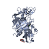
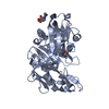
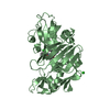
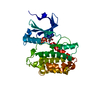
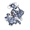
| Title | New Structure Of Cold-Active Protein Tyrosine Phosphatase At 1.1 Angstrom | ||||||
 Components Components | Protein-tyrosine-phosphatase | ||||||
 Keywords Keywords | HYDROLASE / COLD-ACTIVE ENZYME / PSYCHROPHILE / PROTEIN TYROSINE PHOSPHATASE / SHEWANELLA SP. | ||||||
| Function / homology |  Function and homology information Function and homology informationprotein-tyrosine-phosphatase / protein tyrosine phosphatase activity / metal ion binding Similarity search - Function | ||||||
| Biological species |  Shewanella sp. (bacteria) Shewanella sp. (bacteria) | ||||||
| Method |  X-RAY DIFFRACTION / SYNCHROTRON / AB INITIO / Resolution: 1.1 Å X-RAY DIFFRACTION / SYNCHROTRON / AB INITIO / Resolution: 1.1 Å | ||||||
 Authors Authors | Tsuruta, H. / Mikami, B. / Yamamoto, C. / Yamagata, H. | ||||||
 Citation Citation | Journal: Febs J. / Year: 2008 Title: The role of group bulkiness in the catalytic activity of psychrophile cold-active protein tyrosine phosphatase Authors: Tsuruta, H. / Mikami, B. / Yamamoto, C. / Yamagata, H. | ||||||
| History |
|
- Structure visualization
| Structure viewer | Molecule: MolmilJmol/JSmol |
|---|
- Downloads & links
-Download
| PDBx/mmCIF format | 2z72.cif.gz | 188.3 KB | Display | PDBx/mmCIF format |
|---|---|---|---|---|
| PDB format | pdb2z72.ent.gz | 150.8 KB | Display | PDB format |
| PDBx/mmJSON format | 2z72.json.gz | Tree view | PDBx/mmJSON format | |
| Others |  Other downloads Other downloads |
-Validation report
| Arichive directory | https://data.pdbj.org/pub/pdb/validation_reports/z7/2z72ftp://data.pdbj.org/pub/pdb/validation_reports/z7/2z72 | HTTPS FTP |
|---|
-Related structure data
| Related structure data |  2zbmC  1v73S S: Starting model for refinement C: citing same article ( |
|---|---|
| Similar structure data |


-Links
 PDBj
PDBj

- Assembly
| Deposited unit | 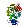
| ||||||||
|---|---|---|---|---|---|---|---|---|---|
| 1 |
| ||||||||
| Unit cell |
|
-Components
| #1: Protein | Mass: 38848.922 Da / Num. of mol.: 1 / Fragment: UNP residues 22-361 Source method: isolated from a genetically manipulated source Source: (gene. exp.) Shewanella sp. (bacteria) / Gene: PPI / Plasmid: PET22B / Production host: | ||||||
|---|---|---|---|---|---|---|---|
| #2: Chemical |   Mass: 65.409 Da / Num. of mol.: 2 / Source method: obtained synthetically / Formula: Zn Mass: 65.409 Da / Num. of mol.: 2 / Source method: obtained synthetically / Formula: Zn#3: Water | ChemComp-HOH / |  Mass: 18.015 Da / Num. of mol.: 772 / Source method: isolated from a natural source / Formula: H2O Mass: 18.015 Da / Num. of mol.: 772 / Source method: isolated from a natural source / Formula: H2OHas protein modification | Y | Sequence details | THERE ARE CONFLICTS BETWEEN SEQRES(SER) AND SEQUENCE DATABASE (GLY). THE AUTHORS BELIEVE THAT THE ...THERE ARE CONFLICTS BETWEEN SEQRES(SER) AND SEQUENCE DATABASE (GLY). THE AUTHORS BELIEVE THAT THE SEQRES IS CORRECT AND IS THE TRUE IDENTITY OF THESE RESIDUES AND IS NATURAL MUTANT. | |
-Experimental details
-Experiment
| Experiment | Method: X-RAY DIFFRACTION |
|---|
- Sample preparation
| Crystal | Density Matthews: 2.23 Å3/Da / Density % sol: 45.08 % |
|---|---|
| Crystal grow | Temperature: 277 K / Method: vapor diffusion, hanging drop / pH: 8.5 Details: 15% polyethyleneglycol, 0.1M ammonium acetate, 50mM p-nitrophenylsulphate, 0.05M Tris-HCl (pH8.5), pH8.50, VAPOR DIFFUSION, HANGING DROP, temperature 277.0K |
-Data collection
| Diffraction | Mean temperature: 103 K |
|---|---|
| Diffraction source | Source: SYNCHROTRON / Site: SPring-8  / Beamline: BL38B1 / Wavelength: 0.7 Å / Beamline: BL38B1 / Wavelength: 0.7 Å |
| Detector | Type: RIGAKU JUPITER 210 / Detector: CCD / Date: Nov 11, 2005 |
| Radiation | Protocol: SINGLE WAVELENGTH / Monochromatic (M) / Laue (L): M / Scattering type: x-ray |
| Radiation wavelength | Wavelength: 0.7 Å / Relative weight: 1 |
| Reflection | Resolution: 1.1→50 Å / Num. obs: 141407 / % possible obs: 89.2 % |
- Processing
| Software |
| |||||||||||||||||||||||||||||||||
|---|---|---|---|---|---|---|---|---|---|---|---|---|---|---|---|---|---|---|---|---|---|---|---|---|---|---|---|---|---|---|---|---|---|---|
| Refinement | Method to determine structure: AB INITIO Starting model: PDB ENTRY 1V73 Resolution: 1.1→8 Å / Num. parameters: 32955 / Num. restraintsaints: 39804 / Cross valid method: FREE R / σ(F): 0 / Stereochemistry target values: ENGH AND HUBER Details: ANISOTROPIC REFINEMENT REDUCED FREE R (NO CUTOFF) BY ?
| |||||||||||||||||||||||||||||||||
| Refine analyze | Num. disordered residues: 39 / Occupancy sum hydrogen: 2670 / Occupancy sum non hydrogen: 3479.15 | |||||||||||||||||||||||||||||||||
| Refinement step | Cycle: LAST / Resolution: 1.1→8 Å
| |||||||||||||||||||||||||||||||||
| Refine LS restraints |
|
