- PDB-5l8o: crystal structure of human FABP6 in complex with cholate -
+
Open data
ID or keywords:
Loading...
-
Basic information
Entry
Database: PDB / ID: 5l8o
Title
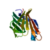
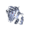
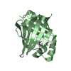
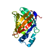
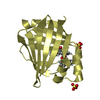
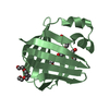
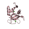
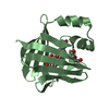
crystal structure of human FABP6 in complex with cholate
Components
Gastrotropin
Keywords
LIPID BINDING PROTEIN / FABP6 / Fatty acid binding protein 6 / Ileal bile acid binding protein / I-BABP / Ileal / Gastrotropin / Fragments / cholate
Function / homology
Function and homology information
NR1H2 & NR1H3 regulate gene expression to control bile acid homeostasis / Triglyceride catabolism / fatty acid transport / Recycling of bile acids and salts / fatty acid binding / lipid metabolic process / negative regulation of cell population proliferation / lipid binding / membrane / nucleus ...NR1H2 & NR1H3 regulate gene expression to control bile acid homeostasis / Triglyceride catabolism / fatty acid transport / Recycling of bile acids and salts / fatty acid binding / lipid metabolic process / negative regulation of cell population proliferation / lipid binding / membrane / nucleus / cytosol / cytoplasm Similarity search - Function
Resolution: 2.39→59.18 Å / Cor.coef. Fo:Fc: 0.939 / Cor.coef. Fo:Fc free: 0.912 / SU B: 11.71 / SU ML: 0.272 / Cross valid method: THROUGHOUT / ESU R: 0.679 / ESU R Free: 0.318 / Details: HYDROGENS HAVE BEEN ADDED IN THE RIDING POSITIONS
Rfactor
Num. reflection
% reflection
Selection details
Rfree
0.28139
731
5.2 %
RANDOM
Rwork
0.22614
-
-
-
obs
0.22896
13374
99.67 %
-
Solvent computation
Ion probe radii: 0.8 Å / Shrinkage radii: 0.8 Å / VDW probe radii: 1.2 Å
 Movie
Movie Controller


 Open data
 Basic information
 Components
 Keywords
 Function and homology information
 Homo sapiens (human)
 X-RAY DIFFRACTION /
  Authors
 Citation
 Structure visualization
 Downloads & links
 Other downloads


 PDBj
PDBj


 Assembly
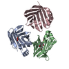


 Mass: 106.120 Da / Num. of mol.: 2 / Source method: obtained synthetically / Formula: C4H10O3
 Mass: 408.571 Da / Num. of mol.: 1 / Source method: obtained synthetically / Formula: C24H40O5
 Mass: 18.015 Da / Num. of mol.: 45 / Source method: isolated from a natural source / Formula: H2O
 Sample preparation
 / Beamline: I02 / Wavelength: 0.9795 Å
 Processing