 Movie
Movie Controller

[English] 日本語
 Yorodumi
- PDB-5l4w: Crystal structure of FimH lectin domain in complex with 3-Fluoro-... -
+ Open data

- Basic information
| Entry | Database: PDB / ID: 5l4w | ||||||
|---|---|---|---|---|---|---|---|
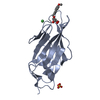
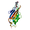
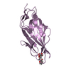
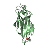
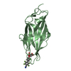
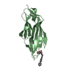
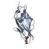
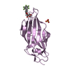
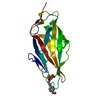
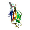
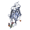
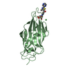
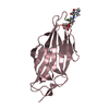
| Title | Crystal structure of FimH lectin domain in complex with 3-Fluoro-Heptylmannoside | ||||||
 Components Components | Protein FimH | ||||||
 Keywords Keywords | SUGAR BINDING PROTEIN / FimH / Type 1 pilus / urinary tract infection / UTI / carbohydrate / lectin / mannose / cell adhesion | ||||||
| Function / homology |  Function and homology information Function and homology informationpilus tip / mechanosensory behavior / Attachment of bacteria to epithelial cells / cell adhesion involved in single-species biofilm formation / pilus / cell-substrate adhesion / D-mannose binding / host cell membrane / cell adhesion Similarity search - Function | ||||||
| Biological species |  | ||||||
| Method |  X-RAY DIFFRACTION / SYNCHROTRON / MOLECULAR REPLACEMENT / Resolution: 1.9 Å X-RAY DIFFRACTION / SYNCHROTRON / MOLECULAR REPLACEMENT / Resolution: 1.9 Å | ||||||
 Authors Authors | Jakob, R.P. / Zihlmann, P. / Rabbani, S. / Maier, T. / Ernst, B. | ||||||
 Citation Citation | Journal: To Be Published Title: High-Affinity Carbohydrate-Lectin Interactions: How Nature Makes it Possible Authors: Zihlmann, P. / Jiang, X. / Sager, C.P. / Fiege, B. / Jakob, R.P. / Siegrist, S. / Zalewski, A. / Rabbani, S. / Eris, D. / Silbermann, M. / Pang, L. / Muhlethaler, T. / Sharpe, T. / Maier, T. / Ernst, B. | ||||||
| History |
|
- Structure visualization
| Structure viewer | Molecule: MolmilJmol/JSmol |
|---|
- Downloads & links
-Download
| PDBx/mmCIF format | 5l4w.cif.gz | 200.8 KB | Display | PDBx/mmCIF format |
|---|---|---|---|---|
| PDB format | pdb5l4w.ent.gz | 162.1 KB | Display | PDB format |
| PDBx/mmJSON format | 5l4w.json.gz | Tree view | PDBx/mmJSON format | |
| Others |  Other downloads Other downloads |
-Validation report
| Arichive directory | https://data.pdbj.org/pub/pdb/validation_reports/l4/5l4wftp://data.pdbj.org/pub/pdb/validation_reports/l4/5l4w | HTTPS FTP |
|---|
-Related structure data
| Related structure data | 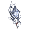 5l4tC  5l4uC 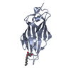 5l4vC 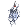 5l4xC 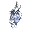 5l4yC 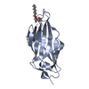 4xo8S S: Starting model for refinement C: citing same article ( |
|---|---|
| Similar structure data |


-Links
 PDBj
PDBj- Assembly
| Deposited unit | 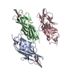
| ||||||||
|---|---|---|---|---|---|---|---|---|---|
| 1 | 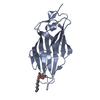
| ||||||||
| 2 | 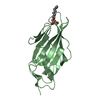
| ||||||||
| 3 | 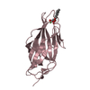
| ||||||||
| Unit cell |
|
-Components
| #1: Protein | Mass: 16916.828 Da / Num. of mol.: 3 Source method: isolated from a genetically manipulated source Source: (gene. exp.) Strain: K12 / Gene: fimH, b4320, JW4283 / Plasmid: pTRC99a / Production host: #2: Sugar |   Type: D-saccharide / Mass: 280.333 Da / Num. of mol.: 3 / Source method: obtained synthetically / Formula: C13H25FO5 Type: D-saccharide / Mass: 280.333 Da / Num. of mol.: 3 / Source method: obtained synthetically / Formula: C13H25FO5#3: Water | ChemComp-HOH / |  Mass: 18.015 Da / Num. of mol.: 603 / Source method: isolated from a natural source / Formula: H2O Mass: 18.015 Da / Num. of mol.: 603 / Source method: isolated from a natural source / Formula: H2O |
|---|
-Experimental details
-Experiment
| Experiment | Method: X-RAY DIFFRACTION / Number of used crystals: 1 |
|---|
- Sample preparation
| Crystal | Density Matthews: 2.87 Å3/Da / Density % sol: 57.16 % |
|---|---|
| Crystal grow | Temperature: 285 K / Method: vapor diffusion, sitting drop Details: 0.2 M (NH4)2SO4, 0.1 M Hepes pH 7 and 25-30% PEG3350 |
-Data collection
| Diffraction | Mean temperature: 100 K |
|---|---|
| Diffraction source | Source: SYNCHROTRON / Site: SLS  / Beamline: X06SA / Wavelength: 1.00001 Å / Beamline: X06SA / Wavelength: 1.00001 Å |
| Detector | Type: DECTRIS PILATUS 6M-F / Detector: PIXEL / Date: Apr 14, 2015 |
| Radiation | Protocol: SINGLE WAVELENGTH / Monochromatic (M) / Laue (L): M / Scattering type: x-ray |
| Radiation wavelength | Wavelength: 1.00001 Å / Relative weight: 1 |
| Reflection | Resolution: 1.9→68.18 Å / Num. obs: 40710 / % possible obs: 90.1 % / Redundancy: 3.5 % / Biso Wilson estimate: 26.67 Å2 / CC1/2: 0.997 / Rmerge(I) obs: 0.101 / Net I/σ(I): 13.8 |
| Reflection shell | Resolution: 1.9→2.01 Å / Redundancy: 3.3 % / Rmerge(I) obs: 0.757 / Mean I/σ(I) obs: 2.2 / % possible all: 71.2 |
- Processing
| Software |
| ||||||||||||||||||||||||||||||||||||||||||||||||||||||||||||||||||||||||||||||||||||||||||||||||||||||||||||||||||
|---|---|---|---|---|---|---|---|---|---|---|---|---|---|---|---|---|---|---|---|---|---|---|---|---|---|---|---|---|---|---|---|---|---|---|---|---|---|---|---|---|---|---|---|---|---|---|---|---|---|---|---|---|---|---|---|---|---|---|---|---|---|---|---|---|---|---|---|---|---|---|---|---|---|---|---|---|---|---|---|---|---|---|---|---|---|---|---|---|---|---|---|---|---|---|---|---|---|---|---|---|---|---|---|---|---|---|---|---|---|---|---|---|---|---|---|
| Refinement | Method to determine structure: MOLECULAR REPLACEMENT Starting model: 4XO8 Resolution: 1.9→68.18 Å / Cor.coef. Fo:Fc: 0.9427 / Cor.coef. Fo:Fc free: 0.9265 / SU R Cruickshank DPI: 0.145 / Cross valid method: THROUGHOUT / σ(F): 0 / SU R Blow DPI: 0.154 / SU Rfree Blow DPI: 0.132 / SU Rfree Cruickshank DPI: 0.128
| ||||||||||||||||||||||||||||||||||||||||||||||||||||||||||||||||||||||||||||||||||||||||||||||||||||||||||||||||||
| Displacement parameters | Biso mean: 27.14 Å2
| ||||||||||||||||||||||||||||||||||||||||||||||||||||||||||||||||||||||||||||||||||||||||||||||||||||||||||||||||||
| Refine analyze | Luzzati coordinate error obs: 0.228 Å | ||||||||||||||||||||||||||||||||||||||||||||||||||||||||||||||||||||||||||||||||||||||||||||||||||||||||||||||||||
| Refinement step | Cycle: LAST / Resolution: 1.9→68.18 Å
| ||||||||||||||||||||||||||||||||||||||||||||||||||||||||||||||||||||||||||||||||||||||||||||||||||||||||||||||||||
| Refine LS restraints |
| ||||||||||||||||||||||||||||||||||||||||||||||||||||||||||||||||||||||||||||||||||||||||||||||||||||||||||||||||||
| LS refinement shell | Resolution: 1.9→1.95 Å / Total num. of bins used: 20
| ||||||||||||||||||||||||||||||||||||||||||||||||||||||||||||||||||||||||||||||||||||||||||||||||||||||||||||||||||
| Refinement TLS params. | Method: refined / Refine-ID: X-RAY DIFFRACTION
| ||||||||||||||||||||||||||||||||||||||||||||||||||||||||||||||||||||||||||||||||||||||||||||||||||||||||||||||||||
| Refinement TLS group |
|
