| Entry | Database: PDB / ID: 5cgb
|
|---|
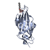
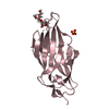
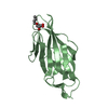
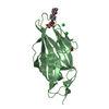
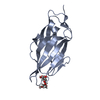
| Title | Crystal structure of FimH in complex with heptyl alpha-D-septanoside |
|---|
 Components Components | Protein FimH |
|---|
 Keywords Keywords | SUGAR BINDING PROTEIN / UTI / lectin / urinary tract infection / type 1 fimbriae / pilus / inhibitor |
|---|
| Function / homology |  Function and homology information Function and homology information
pilus tip / mechanosensory behavior / Attachment of bacteria to epithelial cells / cell adhesion involved in single-species biofilm formation / pilus / cell-substrate adhesion / D-mannose binding / host cell membrane / cell adhesionSimilarity search - Function Fimbrial-type adhesion domain / FimH, mannose-binding domain / FimH, mannose binding / Fimbrial-type adhesion domain / Fimbrial protein / : / Fimbrial-type adhesion domain superfamily / Adhesion domain superfamily / Immunoglobulin-like / Sandwich / Mainly BetaSimilarity search - Domain/homology |
|---|
| Biological species |   Escherichia coli K-12 (bacteria) Escherichia coli K-12 (bacteria) |
|---|
| Method |  X-RAY DIFFRACTION / SYNCHROTRON / MOLECULAR REPLACEMENT / Resolution: 1.6 Å X-RAY DIFFRACTION / SYNCHROTRON / MOLECULAR REPLACEMENT / Resolution: 1.6 Å |
|---|
 Authors Authors | Jakob, R.P. / Preston, R.P. / Zihlmann, P. / Fiege, B. / Sager, C.P. / Vannam, R. / Rabbani, S. / Zalewski, A. / Maier, T. / Ernst, B. / Peczuh, M. |
|---|
 Citation Citation | Journal: Chem Sci / Year: 2018
Title: The price of flexibility - a case study on septanoses as pyranose mimetics.
Authors: Sager, C.P. / Fiege, B. / Zihlmann, P. / Vannam, R. / Rabbani, S. / Jakob, R.P. / Preston, R.C. / Zalewski, A. / Maier, T. / Peczuh, M.W. / Ernst, B. |
|---|
| History | | Deposition | Jul 9, 2015 | Deposition site: RCSB / Processing site: PDBE |
|---|
| Revision 1.0 | Jul 20, 2016 | Provider: repository / Type: Initial release |
|---|
| Revision 1.1 | Apr 7, 2021 | Group: Database references / Category: citation / citation_author
Item: _citation.country / _citation.journal_abbrev ..._citation.country / _citation.journal_abbrev / _citation.journal_id_CSD / _citation.journal_id_ISSN / _citation.journal_volume / _citation.page_first / _citation.page_last / _citation.pdbx_database_id_DOI / _citation.pdbx_database_id_PubMed / _citation.title / _citation.year / _citation_author.identifier_ORCID / _citation_author.name |
|---|
| Revision 1.2 | Jan 10, 2024 | Group: Data collection / Database references / Refinement description
Category: chem_comp_atom / chem_comp_bond ...chem_comp_atom / chem_comp_bond / database_2 / pdbx_initial_refinement_model
Item: _database_2.pdbx_DOI / _database_2.pdbx_database_accession |
|---|
| Revision 1.3 | Nov 13, 2024 | Group: Structure summary / Category: pdbx_entry_details / pdbx_modification_feature |
|---|
|
|---|
 Movie
Movie Controller

 Yorodumi
 Open data
 Basic information
 Structure visualization
 Downloads & links
 Other downloads


 PDBj
PDBj Assembly


 Mass: 292.369 Da / Num. of mol.: 2 / Source method: obtained synthetically / Formula: C14H28O6
 Mass: 96.063 Da / Num. of mol.: 2 / Source method: obtained synthetically / Formula: SO4
 Mass: 18.015 Da / Num. of mol.: 475 / Source method: isolated from a natural source / Formula: H2O
 Sample preparation
 / Beamline: X06SA / Wavelength: 1 Å
 Processing