 Movie
Movie Controller

[English] 日本語
 Yorodumi
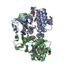
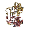
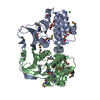
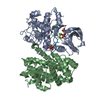
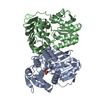
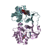
- PDB-5l2w: The X-ray co-crystal structure of human CDK2/CyclinE and Dinaciclib. -
+ Open data

- Basic information
| Entry | Database: PDB / ID: 5l2w | ||||||
|---|---|---|---|---|---|---|---|
| Title | The X-ray co-crystal structure of human CDK2/CyclinE and Dinaciclib. | ||||||
 Components Components |
| ||||||
 Keywords Keywords | Transferase/Cell Cycle/Inhibitor / cyclin-dependent kinase / kinase inhibitor / kinase selectivity / Transferase-Cell Cycle-Inhibitor complex | ||||||
| Function / homology |  Function and homology information Function and homology informationpositive regulation of mesenchymal stem cell proliferation / RHOBTB3 ATPase cycle / cyclin-dependent protein serine/threonine kinase regulator activity / cyclin A1-CDK2 complex / cyclin E2-CDK2 complex / cyclin E1-CDK2 complex / cyclin A2-CDK2 complex / positive regulation of DNA-templated DNA replication initiation / G2 Phase / Y chromosome ...positive regulation of mesenchymal stem cell proliferation / RHOBTB3 ATPase cycle / cyclin-dependent protein serine/threonine kinase regulator activity / cyclin A1-CDK2 complex / cyclin E2-CDK2 complex / cyclin E1-CDK2 complex / cyclin A2-CDK2 complex / positive regulation of DNA-templated DNA replication initiation / G2 Phase / Y chromosome / cyclin-dependent protein kinase activity / regulation of heterochromatin organization / Phosphorylation of proteins involved in G1/S transition by active Cyclin E:Cdk2 complexes / positive regulation of heterochromatin formation / p53-Dependent G1 DNA Damage Response / X chromosome / PTK6 Regulates Cell Cycle / G1/S-Specific Transcription / regulation of anaphase-promoting complex-dependent catabolic process / Defective binding of RB1 mutants to E2F1,(E2F2, E2F3) / centriole replication / Association of TriC/CCT with target proteins during biosynthesis / Regulation of APC/C activators between G1/S and early anaphase / telomere maintenance in response to DNA damage / microtubule organizing center / G0 and Early G1 / centrosome duplication / Telomere Extension By Telomerase / Activation of the pre-replicative complex / cyclin-dependent kinase / cyclin-dependent protein serine/threonine kinase activity / TP53 Regulates Transcription of Genes Involved in G1 Cell Cycle Arrest / positive regulation of G1/S transition of mitotic cell cycle / Regulation of MITF-M-dependent genes involved in cell cycle and proliferation / Activation of ATR in response to replication stress / Cajal body / Cyclin E associated events during G1/S transition / regulation of G2/M transition of mitotic cell cycle / Cyclin A:Cdk2-associated events at S phase entry / cyclin-dependent protein kinase holoenzyme complex / Cyclin A/B1/B2 associated events during G2/M transition / condensed chromosome / mitotic G1 DNA damage checkpoint signaling / cellular response to nitric oxide / negative regulation of protein localization to chromatin / male germ cell nucleus / post-translational protein modification / regulation of mitotic cell cycle / potassium ion transport / cyclin binding / positive regulation of DNA replication / peptidyl-serine phosphorylation / G1/S transition of mitotic cell cycle / meiotic cell cycle / DNA Damage/Telomere Stress Induced Senescence / G2/M transition of mitotic cell cycle / Meiotic recombination / cellular senescence / CDK-mediated phosphorylation and removal of Cdc6 / Transcriptional regulation of granulopoiesis / SCF(Skp2)-mediated degradation of p27/p21 / kinase activity / Orc1 removal from chromatin / Cyclin D associated events in G1 / Regulation of TP53 Degradation / nuclear envelope / Factors involved in megakaryocyte development and platelet production / regulation of gene expression / Processing of DNA double-strand break ends / Senescence-Associated Secretory Phenotype (SASP) / transcription regulator complex / Regulation of TP53 Activity through Phosphorylation / Ras protein signal transduction / protein phosphorylation / chromosome, telomeric region / DNA replication / endosome / chromatin remodeling / protein domain specific binding / protein serine kinase activity / cell division / DNA repair / protein serine/threonine kinase activity / centrosome / positive regulation of cell population proliferation / protein kinase binding / positive regulation of DNA-templated transcription / negative regulation of transcription by RNA polymerase II / magnesium ion binding / signal transduction / DNA-templated transcription / nucleoplasm / ATP binding / nucleus / cytosol / cytoplasm Similarity search - Function | ||||||
| Biological species |  Homo sapiens (human) Homo sapiens (human) | ||||||
| Method |  X-RAY DIFFRACTION / SYNCHROTRON / FOURIER SYNTHESIS / Resolution: 2.8 Å X-RAY DIFFRACTION / SYNCHROTRON / FOURIER SYNTHESIS / Resolution: 2.8 Å | ||||||
 Authors Authors | Chen, P. / Ferre, R.A. / Deihl, W. / Yu, X. / He, Y.-A. | ||||||
 Citation Citation | Journal: Mol.Cancer Ther. / Year: 2016 Title: Spectrum and Degree of CDK Drug Interactions Predicts Clinical Performance. Authors: Chen, P. / Lee, N.V. / Hu, W. / Xu, M. / Ferre, R.A. / Lam, H. / Bergqvist, S. / Solowiej, J. / Diehl, W. / He, Y.A. / Yu, X. / Nagata, A. / VanArsdale, T. / Murray, B.W. | ||||||
| History |
|
- Structure visualization
| Structure viewer | Molecule: MolmilJmol/JSmol |
|---|
- Downloads & links
-Download
| PDBx/mmCIF format | 5l2w.cif.gz | 133.7 KB | Display | PDBx/mmCIF format |
|---|---|---|---|---|
| PDB format | pdb5l2w.ent.gz | 101.4 KB | Display | PDB format |
| PDBx/mmJSON format | 5l2w.json.gz | Tree view | PDBx/mmJSON format | |
| Others |  Other downloads Other downloads |
-Validation report
| Arichive directory | https://data.pdbj.org/pub/pdb/validation_reports/l2/5l2wftp://data.pdbj.org/pub/pdb/validation_reports/l2/5l2w | HTTPS FTP |
|---|
-Related structure data
| Related structure data | 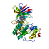 5l2iC  5l2sC 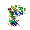 5l2tC 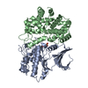 1w98S C: citing same article ( S: Starting model for refinement |
|---|---|
| Similar structure data |


-Links
 PDBj
PDBj


- Assembly
| Deposited unit | 
| ||||||||
|---|---|---|---|---|---|---|---|---|---|
| 1 |
| ||||||||
| Unit cell |
|
-Components
| #1: Protein | Mass: 34143.547 Da / Num. of mol.: 1 Source method: isolated from a genetically manipulated source Source: (gene. exp.) Homo sapiens (human) / Gene: CDK2, CDKN2 / Production host:   Spodoptera frugiperda (fall armyworm) / References: UniProt: P24941, cyclin-dependent kinase Spodoptera frugiperda (fall armyworm) / References: UniProt: P24941, cyclin-dependent kinase |
|---|---|
| #2: Protein | Mass: 35690.262 Da / Num. of mol.: 1 / Fragment: UNP residues 81-363 Source method: isolated from a genetically manipulated source Source: (gene. exp.) Homo sapiens (human) / Gene: CCNE1, CCNE / Production host:  |
| #3: Chemical | ChemComp-1QK / 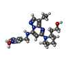  Mass: 397.494 Da / Num. of mol.: 1 / Source method: obtained synthetically / Formula: C21H29N6O2 Mass: 397.494 Da / Num. of mol.: 1 / Source method: obtained synthetically / Formula: C21H29N6O2 |
| #4: Chemical | ChemComp-GOL /   Mass: 92.094 Da / Num. of mol.: 1 / Source method: obtained synthetically / Formula: C3H8O3 Mass: 92.094 Da / Num. of mol.: 1 / Source method: obtained synthetically / Formula: C3H8O3 |
| #5: Water | ChemComp-HOH /  Mass: 18.015 Da / Num. of mol.: 75 / Source method: isolated from a natural source / Formula: H2O Mass: 18.015 Da / Num. of mol.: 75 / Source method: isolated from a natural source / Formula: H2O |
| Has protein modification | Y |
-Experimental details
-Experiment
| Experiment | Method: X-RAY DIFFRACTION / Number of used crystals: 1 |
|---|
- Sample preparation
| Crystal | Density Matthews: 2.73 Å3/Da / Density % sol: 55.03 % |
|---|---|
| Crystal grow | Temperature: 286.15 K / Method: vapor diffusion, sitting drop / Details: PEG20K, 180mM Mg(HCO2)2, 0.1M MES pH 6.0 |
-Data collection
| Diffraction | Mean temperature: 98.15 K |
|---|---|
| Diffraction source | Source: SYNCHROTRON / Site: APS  / Beamline: 17-ID / Wavelength: 1 Å / Beamline: 17-ID / Wavelength: 1 Å |
| Detector | Type: DECTRIS PILATUS3 S 6M / Detector: PIXEL / Date: Nov 9, 2015 |
| Radiation | Protocol: SINGLE WAVELENGTH / Monochromatic (M) / Laue (L): M / Scattering type: x-ray |
| Radiation wavelength | Wavelength: 1 Å / Relative weight: 1 |
| Reflection | Resolution: 2.8→45 Å / Num. obs: 19770 / % possible obs: 99.9 % / Redundancy: 12.76 % / Biso Wilson estimate: 100.25 Å2 / Net I/σ(I): 22.9 |
- Processing
| Software |
| ||||||||||||||||||||||||||||||||||||||||||||||||||||||||||||||||||||||||||||||||||||||||||||||||||||||||||||||||||
|---|---|---|---|---|---|---|---|---|---|---|---|---|---|---|---|---|---|---|---|---|---|---|---|---|---|---|---|---|---|---|---|---|---|---|---|---|---|---|---|---|---|---|---|---|---|---|---|---|---|---|---|---|---|---|---|---|---|---|---|---|---|---|---|---|---|---|---|---|---|---|---|---|---|---|---|---|---|---|---|---|---|---|---|---|---|---|---|---|---|---|---|---|---|---|---|---|---|---|---|---|---|---|---|---|---|---|---|---|---|---|---|---|---|---|---|
| Refinement | Method to determine structure: FOURIER SYNTHESIS Starting model: 1W98 Resolution: 2.8→45 Å / Cor.coef. Fo:Fc: 0.944 / Cor.coef. Fo:Fc free: 0.9244 / SU R Cruickshank DPI: 1.334 / Cross valid method: THROUGHOUT / σ(F): 0 / SU R Blow DPI: 2.166 / SU Rfree Blow DPI: 0.335 / SU Rfree Cruickshank DPI: 0.337
| ||||||||||||||||||||||||||||||||||||||||||||||||||||||||||||||||||||||||||||||||||||||||||||||||||||||||||||||||||
| Displacement parameters | Biso mean: 84.34 Å2
| ||||||||||||||||||||||||||||||||||||||||||||||||||||||||||||||||||||||||||||||||||||||||||||||||||||||||||||||||||
| Refine analyze | Luzzati coordinate error obs: 0.378 Å | ||||||||||||||||||||||||||||||||||||||||||||||||||||||||||||||||||||||||||||||||||||||||||||||||||||||||||||||||||
| Refinement step | Cycle: 1 / Resolution: 2.8→45 Å
| ||||||||||||||||||||||||||||||||||||||||||||||||||||||||||||||||||||||||||||||||||||||||||||||||||||||||||||||||||
| Refine LS restraints |
| ||||||||||||||||||||||||||||||||||||||||||||||||||||||||||||||||||||||||||||||||||||||||||||||||||||||||||||||||||
| LS refinement shell | Resolution: 2.8→2.95 Å / Total num. of bins used: 10
|
