 Movie
Movie Controller

[English] 日本語
 Yorodumi
- PDB-5l2e: Crystal structure of rat Glutamate receptor delta-2 extracellular... -
+ Open data

- Basic information
| Entry | Database: PDB / ID: 5l2e | ||||||
|---|---|---|---|---|---|---|---|
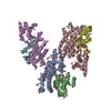
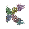
| Title | Crystal structure of rat Glutamate receptor delta-2 extracellular domain | ||||||
 Components Components | Glutamate receptor ionotropic, delta-2,Glutamate receptor ionotropic, delta-2 | ||||||
 Keywords Keywords | PROTEIN BINDING / Synapse Protein / Cell Surface Protein / Glycoprotein / Nervous System | ||||||
| Function / homology |  Function and homology information Function and homology informationtrans-synaptic protein complex / cerebellar granule cell differentiation / positive regulation of long-term synaptic depression / excitatory synapse assembly / synaptic signaling via neuropeptide / regulation of postsynaptic density assembly / positive regulation of synapse assembly / heterophilic cell-cell adhesion / regulation of neuron projection development / AMPA glutamate receptor activity ...trans-synaptic protein complex / cerebellar granule cell differentiation / positive regulation of long-term synaptic depression / excitatory synapse assembly / synaptic signaling via neuropeptide / regulation of postsynaptic density assembly / positive regulation of synapse assembly / heterophilic cell-cell adhesion / regulation of neuron projection development / AMPA glutamate receptor activity / parallel fiber to Purkinje cell synapse / AMPA glutamate receptor complex / ionotropic glutamate receptor complex / regulation of presynapse assembly / regulation of postsynaptic membrane neurotransmitter receptor levels / prepulse inhibition / somatodendritic compartment / regulation of neuron apoptotic process / PDZ domain binding / synaptic transmission, glutamatergic / transmitter-gated monoatomic ion channel activity involved in regulation of postsynaptic membrane potential / excitatory postsynaptic potential / postsynaptic density membrane / modulation of chemical synaptic transmission / intracellular protein localization / scaffold protein binding / dendritic spine / postsynaptic membrane / synapse / glutamatergic synapse / membrane / identical protein binding / plasma membrane Similarity search - Function | ||||||
| Biological species |  | ||||||
| Method |  X-RAY DIFFRACTION / SYNCHROTRON / MOLECULAR REPLACEMENT / Resolution: 4.152 Å X-RAY DIFFRACTION / SYNCHROTRON / MOLECULAR REPLACEMENT / Resolution: 4.152 Å | ||||||
 Authors Authors | Cheng, S. / Ozkan, E. | ||||||
 Citation Citation | Journal: Structure / Year: 2016 Title: Conformational Plasticity in the Transsynaptic Neurexin-Cerebellin-Glutamate Receptor Adhesion Complex. Authors: Cheng, S. / Seven, A.B. / Wang, J. / Skiniotis, G. / Ozkan, E. | ||||||
| History |
|
- Structure visualization
| Structure viewer | Molecule: MolmilJmol/JSmol |
|---|
- Downloads & links
-Download
| PDBx/mmCIF format | 5l2e.cif.gz | 385.3 KB | Display | PDBx/mmCIF format |
|---|---|---|---|---|
| PDB format | pdb5l2e.ent.gz | 314.8 KB | Display | PDB format |
| PDBx/mmJSON format | 5l2e.json.gz | Tree view | PDBx/mmJSON format | |
| Others |  Other downloads Other downloads |
-Validation report
| Arichive directory | https://data.pdbj.org/pub/pdb/validation_reports/l2/5l2eftp://data.pdbj.org/pub/pdb/validation_reports/l2/5l2e | HTTPS FTP |
|---|
-Related structure data
| Related structure data |  5kwrC 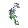 2vt3S  5kc8S S: Starting model for refinement C: citing same article ( |
|---|---|
| Similar structure data |

-Links
 PDBj
PDBj

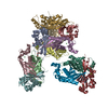
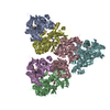
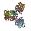
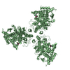
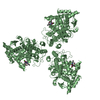
- Assembly
| Deposited unit | 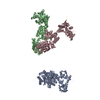
| ||||||||
|---|---|---|---|---|---|---|---|---|---|
| 1 | 
| ||||||||
| 2 | 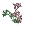
| ||||||||
| Unit cell |
|
-Components
| #1: Protein | Mass: 77833.812 Da / Num. of mol.: 3 Source method: isolated from a genetically manipulated source Source: (gene. exp.)  Trichoplusia ni (cabbage looper) / References: UniProt: Q63226 Trichoplusia ni (cabbage looper) / References: UniProt: Q63226Has protein modification | Y | |
|---|
-Experimental details
-Experiment
| Experiment | Method: X-RAY DIFFRACTION / Number of used crystals: 1 |
|---|
- Sample preparation
| Crystal | Density Matthews: 4.25 Å3/Da / Density % sol: 71.09 % |
|---|---|
| Crystal grow | Temperature: 294 K / Method: vapor diffusion, sitting drop / pH: 6.6 Details: 0.1 M Sodium cacodylate pH 6.6, 1.3 M Ammonium dihydrogen phosphate |
-Data collection
| Diffraction | Mean temperature: 120 K |
|---|---|
| Diffraction source | Source: SYNCHROTRON / Site: APS  / Beamline: 23-ID-D / Wavelength: 1.0332 Å / Beamline: 23-ID-D / Wavelength: 1.0332 Å |
| Detector | Type: DECTRIS PILATUS3 6M / Detector: PIXEL / Date: Apr 19, 2016 |
| Radiation | Monochromator: Double crystal cryo-cooled Si(111) / Protocol: SINGLE WAVELENGTH / Monochromatic (M) / Laue (L): M / Scattering type: x-ray |
| Radiation wavelength | Wavelength: 1.0332 Å / Relative weight: 1 |
| Reflection twin | Operator: -h,-k,l / Fraction: 0.49 |
| Reflection | Resolution: 4.15→50 Å / Num. obs: 30494 / % possible obs: 100 % / Redundancy: 4.2 % / Rmerge(I) obs: 0.157 / Rsym value: 0.157 / Net I/av σ(I): 8.9 / Net I/σ(I): 8.9 |
| Reflection shell | Resolution: 4.15→4.22 Å / Redundancy: 3.9 % / Rmerge(I) obs: 0.795 / Mean I/σ(I) obs: 1.6 / CC1/2: 0.457 / % possible all: 100 |
- Processing
| Software |
| |||||||||||||||||||||||||||||||||||||||||||||||||||||||||||||||||||||||||||||||||||||||||||||||||||||||||
|---|---|---|---|---|---|---|---|---|---|---|---|---|---|---|---|---|---|---|---|---|---|---|---|---|---|---|---|---|---|---|---|---|---|---|---|---|---|---|---|---|---|---|---|---|---|---|---|---|---|---|---|---|---|---|---|---|---|---|---|---|---|---|---|---|---|---|---|---|---|---|---|---|---|---|---|---|---|---|---|---|---|---|---|---|---|---|---|---|---|---|---|---|---|---|---|---|---|---|---|---|---|---|---|---|---|---|
| Refinement | Method to determine structure: MOLECULAR REPLACEMENT Starting model: 2VT3 and 5KC8 Resolution: 4.152→46.583 Å / Cross valid method: FREE R-VALUE / σ(F): 0 / Phase error: 24.5 Details: Refined with twin law -h,-k,l and twin fraction of 0.49.
| |||||||||||||||||||||||||||||||||||||||||||||||||||||||||||||||||||||||||||||||||||||||||||||||||||||||||
| Solvent computation | Shrinkage radii: 0.9 Å / VDW probe radii: 1.11 Å | |||||||||||||||||||||||||||||||||||||||||||||||||||||||||||||||||||||||||||||||||||||||||||||||||||||||||
| Refinement step | Cycle: LAST / Resolution: 4.152→46.583 Å
| |||||||||||||||||||||||||||||||||||||||||||||||||||||||||||||||||||||||||||||||||||||||||||||||||||||||||
| Refine LS restraints |
| |||||||||||||||||||||||||||||||||||||||||||||||||||||||||||||||||||||||||||||||||||||||||||||||||||||||||
| LS refinement shell |
|
