 Movie
Movie Controller

[English] 日本語
 Yorodumi
- PDB-5kwq: Two Tandem RRM Domains of FBP-Interacting Repressor (FIR), also K... -
+ Open data

- Basic information
| Entry | Database: PDB / ID: 5kwq | ||||||
|---|---|---|---|---|---|---|---|
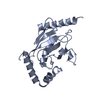
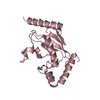
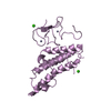
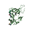
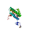
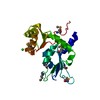
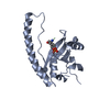
| Title | Two Tandem RRM Domains of FBP-Interacting Repressor (FIR), also Known as PUF60 | ||||||
 Components Components | Poly(U)-binding-splicing factor PUF60 | ||||||
 Keywords Keywords | SPLICING / Tandem RRMs / c-Myc Regulation / Splicing Factor | ||||||
| Function / homology |  Function and homology information Function and homology informationmRNA splice site recognition / alternative mRNA splicing, via spliceosome / mRNA Polyadenylation / regulation of alternative mRNA splicing, via spliceosome / Dengue Virus-Host Interactions / mRNA Splicing - Major Pathway / cadherin binding / ribonucleoprotein complex / apoptotic process / DNA-templated transcription ...mRNA splice site recognition / alternative mRNA splicing, via spliceosome / mRNA Polyadenylation / regulation of alternative mRNA splicing, via spliceosome / Dengue Virus-Host Interactions / mRNA Splicing - Major Pathway / cadherin binding / ribonucleoprotein complex / apoptotic process / DNA-templated transcription / DNA binding / RNA binding / nucleoplasm / identical protein binding Similarity search - Function | ||||||
| Biological species |  Homo sapiens (human) Homo sapiens (human) | ||||||
| Method |  X-RAY DIFFRACTION / MOLECULAR REPLACEMENT / Resolution: 2.8 Å X-RAY DIFFRACTION / MOLECULAR REPLACEMENT / Resolution: 2.8 Å | ||||||
 Authors Authors | Crichlow, G.V. / Yang, Y. / Zhou, H. / Lolis, E.J. / Braddock, D.T. | ||||||
| Funding support |  United States, 1items United States, 1items
| ||||||
 Citation Citation | Journal: Plos One / Year: 2020 Title: Unraveling the mechanism of recognition of the 3' splice site of the adenovirus major late promoter intron by the alternative splicing factor PUF60. Authors: Hsiao, H.T. / Crichlow, G.V. / Murphy, J.W. / Folta-Stogniew, E.J. / Lolis, E.J. / Braddock, D.T. #1: Journal: EMBO J. / Year: 2008Title: Dimerization of FIR upon FUSE DNA binding suggests a mechanism of c-myc inhibition. Authors: Crichlow, G.V. / Zhou, H. / Hsiao, H.H. / Frederick, K.B. / Debrosse, M. / Yang, Y. / Folta-Stogniew, E.J. / Chung, H.J. / Fan, C. / De la Cruz, E.M. / Levens, D. / Lolis, E. / Braddock, D. | ||||||
| History |
|
- Structure visualization
| Structure viewer | Molecule: MolmilJmol/JSmol |
|---|
- Downloads & links
-Download
| PDBx/mmCIF format | 5kwq.cif.gz | 85.1 KB | Display | PDBx/mmCIF format |
|---|---|---|---|---|
| PDB format | pdb5kwq.ent.gz | 62.4 KB | Display | PDB format |
| PDBx/mmJSON format | 5kwq.json.gz | Tree view | PDBx/mmJSON format | |
| Others |  Other downloads Other downloads |
-Validation report
| Arichive directory | https://data.pdbj.org/pub/pdb/validation_reports/kw/5kwqftp://data.pdbj.org/pub/pdb/validation_reports/kw/5kwq | HTTPS FTP |
|---|
-Related structure data
| Related structure data | 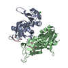 5kvyC 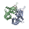 5kw1C 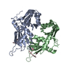 5kw6C  2qfjS S: Starting model for refinement C: citing same article ( |
|---|---|
| Similar structure data |


-Links
 PDBj
PDBj


- Assembly
| Deposited unit | 
| ||||||||
|---|---|---|---|---|---|---|---|---|---|
| 1 | 
| ||||||||
| 2 | 
| ||||||||
| 3 |
| ||||||||
| Unit cell |
|
-Components
| #1: Protein | Mass: 23408.537 Da / Num. of mol.: 2 / Mutation: R106G, C112S, C238A Source method: isolated from a genetically manipulated source Source: (gene. exp.) Homo sapiens (human) / Gene: PUF60, FIR, ROBPI, SIAHBP1 / Plasmid: pET15b / Production host:  |
|---|
-Experimental details
-Experiment
| Experiment | Method: X-RAY DIFFRACTION / Number of used crystals: 1 |
|---|
- Sample preparation
| Crystal | Density Matthews: 1.89 Å3/Da / Density % sol: 34.8 % |
|---|---|
| Crystal grow | Temperature: 293 K / Method: vapor diffusion, hanging drop / pH: 7.5 Details: 1M Lithium sulfate, 0.1M HEPES (pH 7.5), 5% glycerol, mixed with an equal volume of 10 mg/ml protein |
-Data collection
| Diffraction | Mean temperature: 180 K |
|---|---|
| Diffraction source | Source: ROTATING ANODE / Type: RIGAKU MICROMAX-007 / Wavelength: 1.5418 Å |
| Detector | Type: RIGAKU RAXIS IV / Detector: IMAGE PLATE / Date: Mar 13, 2006 / Details: mirrors |
| Radiation | Protocol: SINGLE WAVELENGTH / Monochromatic (M) / Laue (L): M / Scattering type: x-ray |
| Radiation wavelength | Wavelength: 1.5418 Å / Relative weight: 1 |
| Reflection | Resolution: 2.5→50 Å / Num. obs: 11918 / % possible obs: 99.1 % / Observed criterion σ(I): -3 / Redundancy: 2.7 % / Biso Wilson estimate: 110.2 Å2 / Rmerge(I) obs: 0.071 / Net I/av σ(I): 12.7 / Net I/σ(I): 12.3 |
| Reflection shell | Resolution: 2.5→2.59 Å / Redundancy: 2.4 % / Rmerge(I) obs: 0.745 / % possible all: 92.8 |
- Processing
| Software |
| ||||||||||||||||||||||||||||||||||||||||||||||||||||||||||||
|---|---|---|---|---|---|---|---|---|---|---|---|---|---|---|---|---|---|---|---|---|---|---|---|---|---|---|---|---|---|---|---|---|---|---|---|---|---|---|---|---|---|---|---|---|---|---|---|---|---|---|---|---|---|---|---|---|---|---|---|---|---|
| Refinement | Method to determine structure: MOLECULAR REPLACEMENT Starting model: 2QFJ Resolution: 2.8→19.63 Å / Rfactor Rfree error: 0.013 / Data cutoff high absF: 1594738.79 / Data cutoff low absF: 0 / Cross valid method: THROUGHOUT / σ(F): 0
| ||||||||||||||||||||||||||||||||||||||||||||||||||||||||||||
| Solvent computation | Bsol: 36.3683 Å2 / ksol: 0.34409 e/Å3 | ||||||||||||||||||||||||||||||||||||||||||||||||||||||||||||
| Displacement parameters | Biso mean: 49.4 Å2
| ||||||||||||||||||||||||||||||||||||||||||||||||||||||||||||
| Refine analyze |
| ||||||||||||||||||||||||||||||||||||||||||||||||||||||||||||
| Refinement step | Cycle: 1 / Resolution: 2.8→19.63 Å
| ||||||||||||||||||||||||||||||||||||||||||||||||||||||||||||
| Refine LS restraints |
| ||||||||||||||||||||||||||||||||||||||||||||||||||||||||||||
| LS refinement shell | Resolution: 2.8→2.98 Å / Rfactor Rfree error: 0.038 / Total num. of bins used: 6
|
