 Movie
Movie Controller

[English] 日本語
 Yorodumi
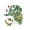
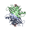
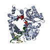
- PDB-5kvy: CRYSTAL STRUCTURE OF THE TWO TANDEM RRM DOMAINS OF PUF60 BOUND TO... -
+ Open data

- Basic information
| Entry | Database: PDB / ID: 5kvy | |||||||||
|---|---|---|---|---|---|---|---|---|---|---|
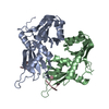
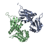
| Title | CRYSTAL STRUCTURE OF THE TWO TANDEM RRM DOMAINS OF PUF60 BOUND TO A PORTION OF AN ADML PRE-MRNA 3' SPLICE SITE ANALOG | |||||||||
 Components Components |
| |||||||||
 Keywords Keywords | SPLICING/DNA / TANDEM RRMS / PROTEIN-NUCLEIC ACID COMPLEX / SPLICING FACTOR / SPLICING-DNA complex | |||||||||
| Function / homology |  Function and homology information Function and homology informationmRNA splice site recognition / alternative mRNA splicing, via spliceosome / regulation of alternative mRNA splicing, via spliceosome / mRNA Splicing - Major Pathway / cell junction / cadherin binding / ribonucleoprotein complex / apoptotic process / DNA binding / RNA binding ...mRNA splice site recognition / alternative mRNA splicing, via spliceosome / regulation of alternative mRNA splicing, via spliceosome / mRNA Splicing - Major Pathway / cell junction / cadherin binding / ribonucleoprotein complex / apoptotic process / DNA binding / RNA binding / nucleoplasm / identical protein binding Similarity search - Function | |||||||||
| Biological species |  Homo sapiens (human) Homo sapiens (human)synthetic construct (others) | |||||||||
| Method |  X-RAY DIFFRACTION / SYNCHROTRON / MOLECULAR REPLACEMENT / molecular replacement / Resolution: 1.95 Å X-RAY DIFFRACTION / SYNCHROTRON / MOLECULAR REPLACEMENT / molecular replacement / Resolution: 1.95 Å | |||||||||
| Model details | protein / nucleic acid complex | |||||||||
 Authors Authors | Hsiao, H.-H. / Crichlow, G.V. / Albright, R.A. / Murphy, J.W. / Lolis, E.J. / Braddock, D.T. | |||||||||
| Funding support |  United States, 1items United States, 1items
| |||||||||
 Citation Citation | Journal: Plos One / Year: 2020 Title: Unraveling the mechanism of recognition of the 3' splice site of the adenovirus major late promoter intron by the alternative splicing factor PUF60. Authors: Hsiao, H.T. / Crichlow, G.V. / Murphy, J.W. / Folta-Stogniew, E.J. / Lolis, E.J. / Braddock, D.T. #1: Journal: EMBO J. / Year: 2008Title: Dimerization of FIR upon FUSE DNA binding suggests a mechanism of c-myc inhibition Authors: Crichlow, G.V. / Zhou, H. / Hsiao, H.H. / Frederick, K.B. / Debrosse, M. / Yang, Y. / Folta-Stogniew, E.J. / Chung, H.J. / Fan, C. / De la Cruz, E.M. / Levens, D. / Lolis, E. / Braddock, D. | |||||||||
| History |
|
- Structure visualization
| Structure viewer | Molecule: MolmilJmol/JSmol |
|---|
- Downloads & links
-Download
| PDBx/mmCIF format | 5kvy.cif.gz | 99 KB | Display | PDBx/mmCIF format |
|---|---|---|---|---|
| PDB format | pdb5kvy.ent.gz | 71.3 KB | Display | PDB format |
| PDBx/mmJSON format | 5kvy.json.gz | Tree view | PDBx/mmJSON format | |
| Others |  Other downloads Other downloads |
-Validation report
| Arichive directory | https://data.pdbj.org/pub/pdb/validation_reports/kv/5kvyftp://data.pdbj.org/pub/pdb/validation_reports/kv/5kvy | HTTPS FTP |
|---|
-Related structure data
| Related structure data | 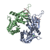 5kw1C  5kw6C 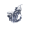 5kwqC  2qfjS C: citing same article ( S: Starting model for refinement |
|---|---|
| Similar structure data |


-Links
 PDBj
PDBj


- Assembly
| Deposited unit | 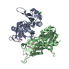
| ||||||||
|---|---|---|---|---|---|---|---|---|---|
| 1 |
| ||||||||
| Unit cell |
| ||||||||
| Details | Authors have supported the biological unit analysis by performing size exclusion chromatography and light scattering |
-Components
| #1: Protein | Mass: 23408.537 Da / Num. of mol.: 2 / Fragment: tandem RRM domains / Mutation: R123G, C129S, C255A Source method: isolated from a genetically manipulated source Source: (gene. exp.) Homo sapiens (human) / Gene: PUF60, FIR, ROBPI, SIAHBP1 / Plasmid: pET15b / Production host:  #2: DNA chain | | Mass: 8862.427 Da / Num. of mol.: 1 / Fragment: AdML3' / Source method: obtained synthetically Details: Sequence from the adenovirus major late pre-mRNA 3'splice site Source: (synth.) synthetic construct (others) #3: Chemical | ChemComp-CL / |   Mass: 35.453 Da / Num. of mol.: 1 / Source method: obtained synthetically / Formula: Cl Mass: 35.453 Da / Num. of mol.: 1 / Source method: obtained synthetically / Formula: Cl#4: Water | ChemComp-HOH / |  Mass: 18.015 Da / Num. of mol.: 219 / Source method: isolated from a natural source / Formula: H2O Mass: 18.015 Da / Num. of mol.: 219 / Source method: isolated from a natural source / Formula: H2O |
|---|
-Experimental details
-Experiment
| Experiment | Method: X-RAY DIFFRACTION / Number of used crystals: 1 |
|---|
- Sample preparation
| Crystal | Density Matthews: 1.68 Å3/Da / Density % sol: 26.93 % |
|---|---|
| Crystal grow | Temperature: 293 K / Method: vapor diffusion, hanging drop / pH: 8.7 Details: 0.1 M TRIS-HCl, 25% PEG 4000, 5-10 mM Barium Chloride Dihydrate, pH 8.7, mixed with 10 mg/ml protein-nucleic acid mixture in 50 mM TRIS-HCl, pH 8.0, 150 mM NaCl, 20 micromolar EDTA. |
-Data collection
| Diffraction | Mean temperature: 100 K |
|---|---|
| Diffraction source | Source: SYNCHROTRON / Site: APS / Beamline: 24-ID-C / Wavelength: 0.9795 Å |
| Detector | Type: ADSC QUANTUM 315 / Detector: CCD / Date: Jul 9, 2009 |
| Radiation | Monochromator: SI(111) / Protocol: SINGLE WAVELENGTH / Monochromatic (M) / Laue (L): M / Scattering type: x-ray |
| Radiation wavelength | Wavelength: 0.9795 Å / Relative weight: 1 |
| Reflection | Resolution: 1.95→30 Å / Num. obs: 26419 / % possible obs: 99.7 % / Observed criterion σ(I): -3 / Rmerge(I) obs: 0.059 |
| Reflection shell | Resolution: 1.95→2 Å / Rmerge(I) obs: 0.452 / Mean I/σ(I) obs: 2.2 / % possible all: 100 |
-Phasing
| Phasing | Method: molecular replacement |
|---|
- Processing
| Software |
| ||||||||||||||||||||||||||||||||||||||||||||||||||||||||||||||||||||||||||||||||
|---|---|---|---|---|---|---|---|---|---|---|---|---|---|---|---|---|---|---|---|---|---|---|---|---|---|---|---|---|---|---|---|---|---|---|---|---|---|---|---|---|---|---|---|---|---|---|---|---|---|---|---|---|---|---|---|---|---|---|---|---|---|---|---|---|---|---|---|---|---|---|---|---|---|---|---|---|---|---|---|---|---|
| Refinement | Method to determine structure: MOLECULAR REPLACEMENT Starting model: 2QFJ Resolution: 1.95→30 Å / Cross valid method: THROUGHOUT / Details: DATA WERE DE-TWINNED DURING REFINEMENT PROCESS
| ||||||||||||||||||||||||||||||||||||||||||||||||||||||||||||||||||||||||||||||||
| Displacement parameters | Biso mean: 34.49 Å2
| ||||||||||||||||||||||||||||||||||||||||||||||||||||||||||||||||||||||||||||||||
| Refine analyze |
| ||||||||||||||||||||||||||||||||||||||||||||||||||||||||||||||||||||||||||||||||
| Refinement step | Cycle: LAST / Resolution: 1.95→30 Å
| ||||||||||||||||||||||||||||||||||||||||||||||||||||||||||||||||||||||||||||||||
| Refine LS restraints |
| ||||||||||||||||||||||||||||||||||||||||||||||||||||||||||||||||||||||||||||||||
| LS refinement shell | Resolution: 1.95→2.02 Å
|
